Chapter: 11th 12th standard bio zoology Human Body higher secondary school
Anatomy of Cycas
Cycas
Division : Cycadophyta
Class : Cycadopsida
Order : Cycadales
Family : Cycadaceae
Genus : Cycas
Gymnosperms are plants which produce naked seeds i.e., plants which lack ovary and hence do not produce fruits. Cycas belongs to this group of plants.
The genus cycas is the most widely distributed genus of the order cycadales. There are about 20 species which grow in the wilderness in China, Japan, Australia, Africa, Nepal, Bangladesh, Burma and India. C. circinalis, C. pectinata, C. rumphii and C. beddomei, are found in the wilderness in India. C. revoluta is grown in gardens in India.
Species of Cycas are of considerable economic importance. Starch is extracted from several species of cycas. Young succulent leaves are used as vegetable in some parts of India. Several species of cycas are of medicinal value. The juice of young leaves of C. circinalis is used as a remedy for stomach disorders, flatulence, blood vomiting and skin diseases. The decoction of young seeds of this species is purgative and emetic. A tincture prepared from the seeds of C. revoluta is used to relieve headache, giddiness and sore throat.
Anatomy
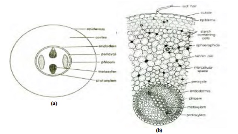
Normal root: A cross section of normal root (fig) consists of epiblema, cortex and central vascular tissue.
Epiblema: It is composed of a single layer of thin-walled cells.
Cortex: It is a multilayered zone of thin-walled parenchymatous cells. These cells are filled with starch. Tannin cells and mucilage cells are also present in the cortex. The innermost layer of the cortex is endodermis. Pericycle is a multi-layered zone found next to endodermis.
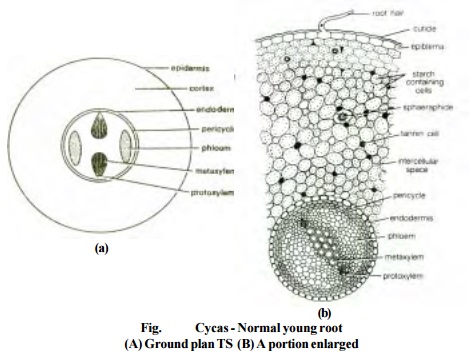
Vascular tissue: This tissue forms a central diarch stele. The diarch steel refers to the presence of two patches of protoxylem points. The xylem consists of xylem tracheids. A tracheid is one celled, non-living, elongated xylem element with thick lignified and pitted cell walls. The xylem is exarch i.e., the protoxylem is pointing towards the periphery while the metaxylem is located near the centre of roots. The pith is either reduced or completely absent.
The normal roots exhibit secondary growth, which starts by the formation of cambium strips that are formed inner to the primary phloem strands (fig). These cambium strips cut off secondary phloem towards the outer side and secondary xylem towards the inner side. Due to the development of secondary structures the primary phloem is crushed while the primary xylem is found in the centre. A distinct layer of cork cambium (phellogen) arisis in the outer region of cortex which gives rise to cork (phellem) on its outer side and secondary cortex (phelloderm) on its inner side. Cork, cork cambium and cork cortex or secondary cortex are collectively known as periderm.
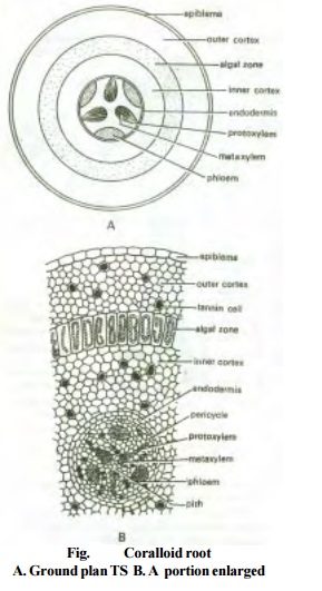
Coralloid roots: The internal structure of coralloid roots is similar to that of normal roots except in certain respects. The cortex of coralloid root is differentiated into i) outer cortex composed of polygonal cells, ii) inner cortex consisted of thin-walled parenchymatous cells and iii) middle cortex made up of a single layer of loosely connected thin-walled and radially elongated cells with blue green algal forms such as Anabaena or Nostoc. Coralloid roots show little or no secondary growth.
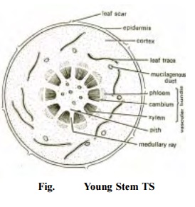
Stem : The stem is irregular in outline due to the presence of numerous persistent leaf bases. Its internal structure is similar to that of dicots of Angiosperms. Young stem of cycas is differentiated into epidermis, cortex and vascular cylinder (fig). The epidermis
is the outermost layer of stem covered with a thick cuticle. Cortex forms the major part of the stem. It is composed of parenchymatous cells with rich starch grains. The cortex is traversed by several mucilagenous canals and many leaf traces. The inner most layer of cortex is endodermis which is followed by pericycle. However, these two regions are not distinctly seen.
In the young stem, vascular region is very small when compared to the cortical zone. There are several vascular bundles arranged in a ring. The vascular bundles are conjoint, collateral, endarch and open. The individual bundles are separated by parenchymatous medullary rays. The xylem consists of tracheids and paraenchyma. Xylem vessels are absent. The phloem consists of sieve tubes and phloem parenchyma. There are no companion cells.
There is a parenchymatous pith present in the centre of the stem. The pith cells are rich in starch and some cells contain tannin and mucilagenous substances.
Secondary growth i.e., the formation of secondary xylem and secondary phloem from cambium as found in dicot stems, is observed in old stems of cycas. In addition to secondary xylem and secondary phloem, the cambium also forms parenchymatous medullary rays. A well developed stem of cycas is called manoxylic because the wood is not compact due to well developed pith, cortex and broad medullary rays with limited vasculature.
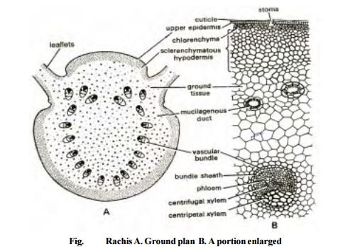
Rachis : Transverse section of rachis is more or less circular in outline. It has two rows of leaflets inserted on one side. The internal structure is differentiated into epidermis, hypodermis, ground tissue and vascular tissue (fig).
Epidermis is covered with a thick cuticle. The epidermis is interrupted by sunken stomata. The epidermis is followed by hypodermis. The hypodermis consists of outer thin-walled chlorenchymatous cells (2-3 layers) and the inner thick walled sclerenchymatous cells (4-5 layers). The ground tissue consists of parenchymatous cells with mucilage canals.
The vascular bundles are arranged in an inverted omega-shaped manner. The bundles are conjoint, collateral, open and diploxylic. Diploxylic condition refers to the presence of centrifugal and centripetal xylem.
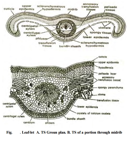
Leaflet : Transverse section of cycas leaflet shows the following tissues i) upper and lower epidermis, ii) hypodermis, iii) mesophyll, iv) transfusion tissue and v) vascular bundles.
1. The upper and lower epidermis are the outermost cellular layers (one celled thick) of the upper and lower sides respectively of the leaflets. Both of them are covered by thick cuticle. The upper epidermis is continuous, whereas the lower epidermis is interrupted by sunken stomata.
2. Hypodermis : This layer is made up of sclerenchymatous cells. The hypodermal layer protects the plant from over-heating and excessive transpiration.
3. Mesophyll : This tissue consists of palisade and spongy parenchyma cells. The palisade layer is a single continuous layer of column-like cells. The spongy parenchyma consists of several layers of loosely arranged oval or irregular cells. Both pallisade and spongy parenchyma cells are rich in chloroplasts.
4. Transfusion tissue : This tissue consists of two small groups of short and wide tracheid-like cells with thickenings / pits on their walls. A few layers of transversely
5. elongated cells are present in both the wings between palisade and spongy parenchyma cells. These layers are called accessory transfusion tissue or secondary transfusion tissue.
6. Vascular bundle : There is only one vascular bundle present in the midrib region of the leaflet. It is conjoint, collateral, open and diploxylic. The triangular centrifugal xylem is well-developed with endarch protoxylem. Phloem is arc-shaped and remains separated by cambium. Phloem consists of sieve tubes and phloem parenchyma. Companion cells are absent.
Related Topics