Chapter: Biology: Practical Zoology
Toad: External Features, Digestive System, Method of Dissection, Skeleton
Toad
Toad: Toad is a familiar animal of the class Amphibia. Its body is bilaterally symmetrical. The body of toad is gray coloured with warts throughout and divided into two portions, namely:
1.Head 2.Trunk
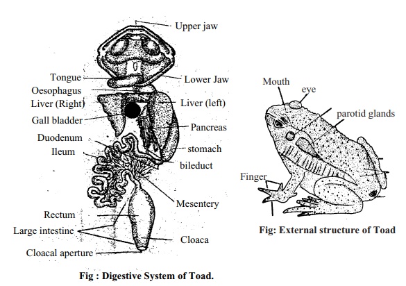
1. Head: The shape of head is like an equilateral triangle. Its mouth is verylarge and bordered by jaws. In front of the head there are nasal apertures and at the back two eyes. Behind the eyes there are two tympanic membrane's. Two glands known as parotid glands are located behind the tympanum.
2. Trunk: The portion of body extending from fore limbs to hind limbs isknown as trunk. At the end of trunk lies the anus.
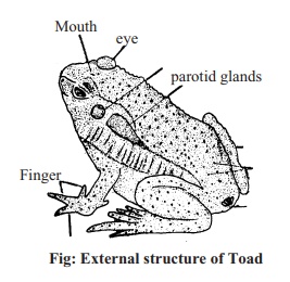
Digestive system of Toad:
The digestive system consists of:
1. Alimentary Canal
2. Digestive Glands.
1. Alimentary Canal: Alimentar Canal of toad is tube like and extended from the mouth to the anus. It consists of the following organs. e.g.
Mouth: The mouth and the buccal cavity is more or less wide and covered by toothless jaws. Upper jaw is fixed.
Buccal Cavity : It is situated just behind the mouth opening. On the anterior end of the lower jaw there islong fleshy tongue which is bluntanteriorlyThe opening behind the tongue is glottis.
Pharynx: It is the last part of buccal cavity.
Oesophagus: In the lower part of pharynx there is a narrow tube known asoesophagus.
Stomach: It is sac-like and fleshy, wider anteriorly and narrowerposteriorly. Its wider end is the cardiac end and narrow posterior end is pyloric end. This end is connected with small intestine.
Small Intestine: It has two portions, namely: a. Duodenum b. Ileum.
a. Duodenum: It is the anterior narrow portion, connected with the stomach.It forms U-shaped structure.
b. Ilium: It is extended from duodenum to large intestine and spirally coiled.
Large Intestine: It is a short and wider portion of intestine. The wider partis the rectum and the narrower part is cloaca.
Cloacal Opening: It is at the terminal end of digestive system throughwhich faeces and urine are eliminated to the exterior.
2. Digestive Glands:
Liver: It is the largest gland in thebody of toad. It is divided into two portions. On either side of the heart the liver is situated. At the mid region of the liver the gall-bladder is located. Pancreas: In between the duodenum and ileum lies the pancreas which is light yellowish in colour.
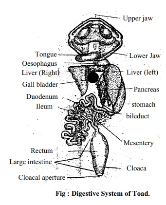
Method of Dissection:
Bring a live toad in the laboratory, keep it in a glass jar. Insert cotton wool soaked with chloroform in the jar for a few minutes. The toad will be anaesthetized.
If chloroform in not available, push a pin in the brain or feed the toad saline water to senseless it. Now place the toad on the dissecting tray keeping the ventral side upward. Pull the fore limbs and hind limbs straight and fixed them with the pins in wax tray. Now pour water in the tray.
Now hold the skin with the forceps and dissect to separate the skin from the fleshy part of the body. Pin up the skin on two sides in the tray.
Now observe the different parts of the alimentary system. according to the figure.
Equipment: The instruments required for dissection of toad are wax-traychloroform, forceps, blade or scissor and pins.
Skeleton of toad :
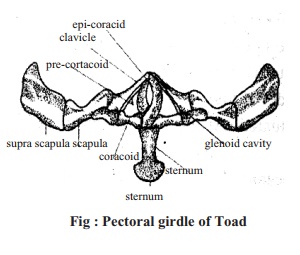
Pectoral Girdle:
a) The Pectoral girdle consists of two similar and symmetrical half.
b) Each half consists of suprascapula, scapula, clavicle, coracoid, precoracoid, glenoid cavity and epicoraciod.
c) Sternum is located at the lower end of pectoral girdle.
d) Suprascapula is wide and thin. Scapula is hard thick and elongated.
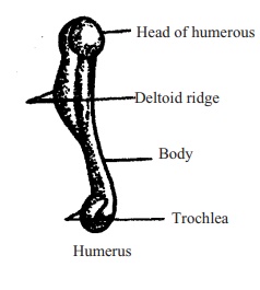
Humerus : It is the first bone of forelimb of toad.
a) This bone is long and narrow towards the posterior end.
b) At the tip, there is a round head like structure.
c) The bone has a trochlea at the posterior end.
d) Deltoid ridge is present.
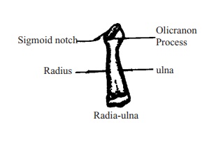
Radia-ulna: It is the radia-ulna offorelimb of toad.
· The two bones radius and ulna, being fused together to form the bone.
· The posterior end of this bone is comparatively wide.
· The anterior end has a concave sigmoid notch.
Carpals, Metacarpals and Phalanges:
a. Carpals:
Next to radia-ulna there are small round bones known as carpal.
Bones are arranged in two rows.
b. Metacarpals:
They are next to carpal.
Four narrow long bones form the metacarpal.
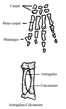
c. Phalanges:
l. There are four phalanges in the forelimb.
1st and 2nd finger have two phalanges.
3rd and 4th finger have three phalanges.
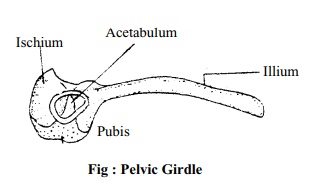
Pelvic Girdle:
1. It is like the English letter "V'.
2. The girdle is divided into two similar parts. Pubis
3. In each half there are illium, ischium and pubis. Fig : Pelvic Girdle
4 In the junction of these three bones there is acetabulum.
Femur: It is the first bone of the hindlimb of toad.
The bone is elongated and sticklike.
The bone is a little curved.
The tip of the bone is round. Posterior end has a swelling.
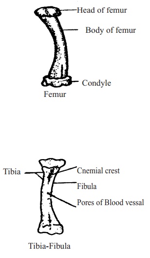
Tibia-Fibula:
It is the tibia-fibula of hind leg of toad.
1.The bone is long and the ends are wide. Tibia
2. Two bones Tibia and Fibula being fused together form the bone, at the junction there is a cnemial crest.
3.The elevated regions on both sides of the depression are clearly seen.
Astragalus and Calcaneum: It is the bone of hind leg of toad.
a) It is made of two bones astragalus and calcaneum.
b) Both the bones are narrow elongated with a gap in-between.
c) The terminal ends of both the bones are fused.
d) Cartilage is present at the two ends.
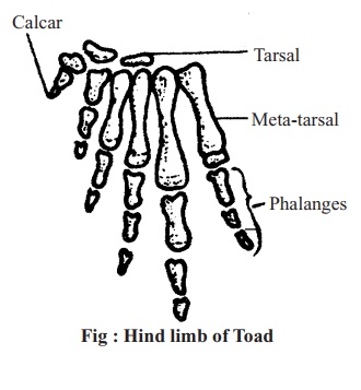
Tarsals : The Tarsal bones are arranged intwo rows. 1st row is formed by two small bones. And these two bones are just belowthe astragalus and calcaneum.
Metatarsals: Five narrow and long bonesform this metatarsal.
Phalanges:
1. These are finger bones.
2. There are five fingers in the hind limb of Toad.
3. There are three phalanges in 1st, 2nd and 5th finger and four phalanges in 4th finger.
First Vertebra or Atlas: It is the first vertebra of the vertebral column of Toad.
1. It is ring like in appearance.
2. Transverse process is absent.
3. Neural canal is big.
4. Centrum is small but wide.
5. Prezygapophysis is absent.
6. Front end of centrum is concave and posterior end is convex.
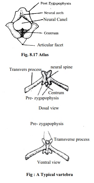
Typical Vertebra:
It is the typical vertebra of Toad.
Small, thick centrum is present.
Small and blunt neural spine present.
On both sides of neural arch there is transverse process.
Prezygapophysis and postzygapohsis are present.
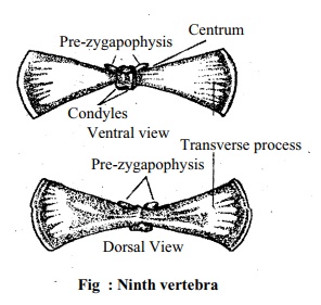
Ninth Vertebra: It is the ninth vertebra of the vertebral column of Toad.
Transverse process is wide.
At the anterior end there is centrum.
On the posterior region there are two condyles.
Prezygapophysis is present but postzygapophysis is absent.
Neural canal and neural arch are not completely developed.
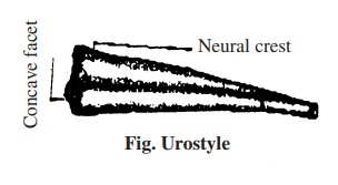
Urostyle: It is the last bone of vertebral.
column of toad or urostyle.
1. Urostyle looks like a rod.
2. It gradually narrows posteriorly.
3. At the anterior end there is concave depression or facet.
4. At the midline of the bone there is a neural crest like a blade of a knife.
5. Neural canal is very narrow.
Related Topics