Chapter: Biochemical Pharmacology : The ionic basis of cell excitation
The origin of cell excitation
The origin of cell excitation
The mechanisms we have
discussed above account for the propagation and for the termination of the
action potential. However, so far we have relied on external electrodes for its
initiation. Under physiological conditions, action potentials can be evoked in
various ways.
The first, very important means of action
potential genera-tion consists in synaptic transmission. A synapse connects a
presynaptic cell (always a neuron) to a postsynaptic cell (a neuron or muscle
cell). In brief, a synapse works as fol-lows:
1. Excitation of the presynaptic cell leads to the
release of a neurotransmitter substance.
2. The neurotransmitter binds to a receptor on the
postsy-naptic cell, very commonly a ligand-gated channel.
3. The receptor channel opens and locally
depolarizes the membrane.
4. The local depolarization is picked up by
adjacent voltage-gated channels and propagated across the entire membrane of
the postsynaptic cell.
A very widespread receptor
channel is the nicotinic acetyl-choline receptor, which is found on all
skeletal muscle cells. Upon binding of the transmitter (acetylcholine), this
chan-nel opens up to both K+ and Na+. This would drag the
mem-brane potential towards the mean value between the two ions' equilibrium
potentials (Figure 4.10). In the process, however, the firing level of the
postsynaptic membrane is reached (see Figure 4.6), the adjacent voltage-gated
sodium channels open, and the action potential starts propagating along the
postsynaptic membrane in the usual way. We will see more about synapses in a
later chapter.
Another principal means of
action potential generation con-sists in spontaneous, rhythmic membrane
depolarization. This occurs in specialized pacemaker cells in heart and smooth
muscle. Therefore, while these tissues are modulat-ed by neuronal and hormonal
influences, they are capable of self-stimulation in the absence of any neuronal
control.
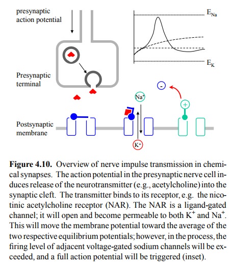
Figure 4.10. Overview of nerve impulse transmission in chemi-cal synapses. The
action potential in the presynaptic nerve cell in-duces release of the
neurotransmitter (e.g., acetylcholine) into the synaptic cleft. The transmitter
binds to its receptor, e.g. the nico-tinic acetylcholine receptor (NAR). The
NAR is a ligand-gated channel; it will open and become permeable to both K+
and Na+. This will move the membrane potential toward the average of
the two respective equilibrium potentials; however, in the process, the firing
level of adjacent voltage-gated sodium channels will be ex-ceeded, and a full
action potential will be triggered (inset).
There are two major
differences between action potentials that occur in nerve cells or skeletal
muscle cells on the one hand, and in heart muscle cells on the other:
1. The duration of the action potential in the
heart is much longer – several hundred milliseconds as opposed to sev-eral
milliseconds in nerve and skeletal muscle. While each skeletal muscle
contraction is triggered and sus-tained by a repetitive burst of many action
potentials, in the heart there is only one action potential per heart beat.
2. While sodium is the major ion species
responsible for excitation in nerve cells and skeletal muscle, in the heart
pacemaker cells this role is taken by calcium. Calcium also has a prominent
role in the excitation of smooth muscle cells.
Two types of calcium channels
control the spontaneous for-mation of an action potential. These channels
differ in their respective response to the prevailing membrane potential. One
of them (the CaT channel) opens slowly but steadily at low
potentials, thereby ramping up the membrane potential to the firing level. At
this point, the Ca L channel responds and induces rapid and complete
membrane depolarization (Figure 4.11).
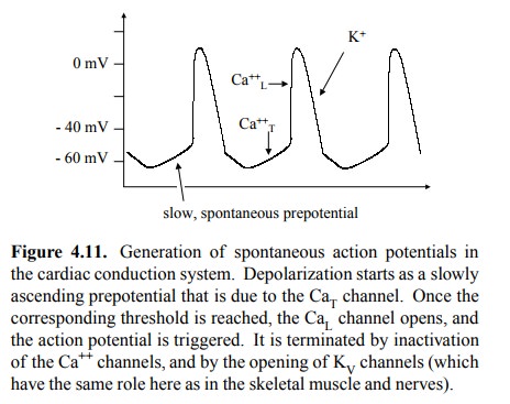
Figure 4.11. Generation of spontaneous action potentials in the cardiac conduction system. Depolarization starts as a slowly
ascending prepotential that is due to the CaT channel. Once the
corresponding threshold is reached, the CaL channel opens, and the
action potential is triggered. It is terminated by inactivation of the Ca++
channels, and by the opening of KV channels (which have the same role
here as in the skeletal muscle and nerves).
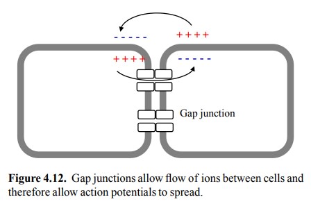
The heart also provides us
with the `classical' example of the third major way to trigger an action
potential, which is by electrical coupling to a neighbouring cell via gap
junc-tions (Figure 4.12). The excitation that is spontaneously generated in the
small number of specialized pacemaker cells in the conduction system5
spreads in this way across the entire heart and ensures coordinated action. The
speed of conduction varies in different parts of the heart, and the atria are
excited and will contract before the ventricles. Groups of smooth muscle cells
in many organs are likewise connected to each other and thus behave as
functional units in a similar way.
Related Topics