Chapter: Clinical Dermatology: Diagnosis of skin disorders
The ichthyoses - Disorders of keratinization
The
ichthyoses
The
word ichthyosis comes from the Greek word for a fish. It is applied to
disorders that share, as their main feature, a dry rough skin with marked
scaling but no inflammation. Strictly speaking, the scales lack the regular
overlapping pattern of fish scales, but the term is usefully descriptive and
too well entrenched to be discarded. There are several types
Ichthyosis vulgaris
Cause
Inherited
as an autosomal dominant disorder, this condition is common and affects about 1
person in The relevant gene may be concerned with the production of
profilaggrin, a precursor of filaggrin, itself a component of keratohyalin
granules.
Presentation
The
dryness is usually mild and symptoms are few. The scales are small and branny,
being most obvious on the limbs and least obvious in the major flexures. The
skin creases of the palm may be accentuated. Keratosis pilaris is often present on the limbs.
Clinical course
The
skin changes are not usually present at birth but develop over the first few
years of life. Some patients improve in adult life, particularly during warm
weather, but the condition seldom clears completely.
Complications
The
already dry skin chaps in the winter and is easily irritated by degreasing
agents. This should be taken into account in the choice of a career. Ichthyosis
of this type is apt to appear in a stubborn combination with atopic eczema.
Differential diagnosis
It
can usually be distinguished from less common types of ichthyosis on the basis
of the pattern of inheritance and of the type and distribution of the scaling.
Investigations
None
are usually needed.
Treatment
This
is palliative. The dryness can be helped by the regular use of emollients,
which are best applied after a shower or bath. Emulsifying ointment, soft white
paraffin, E45 and unguentum merck are all quite suit-able (Formulary 1) and the
selection depends on the patientŌĆÖs preference. Many find proprietary bath oils
and creams containing urea or lactic acid helpful also (Formulary 1).
X-linked recessive ichthyosis
Cause
This
less common type of ichthyosis is inherited as an X-linked recessive trait and
therefore, in its complete form, is seen only in males, although some female
carriers show mild scaling. The condition affects about 1 in 6000 males in the
UK and is associated with a deficiency of the enzyme steroid sulphatase, which
hydrolyses cholesterol sulphate. The responsible gene has been localized to the
terminal part of the X chro-mosome at Xp 22.3.
Presentation and course
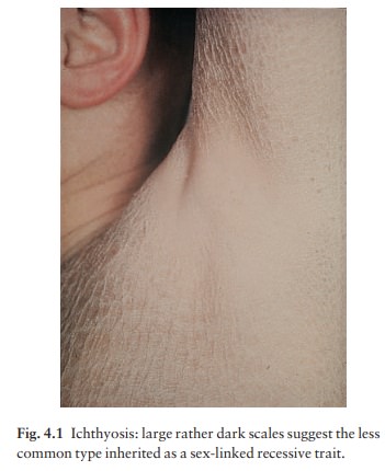
In
contrast to the delayed onset of the dominantly inherited ichthyosis vulgaris,
scaling appears early, often soon after birth, and always by the first
birth-day. The scales are larger and browner (Fig. 4.1), involve the neck, and
to a lesser extent the popliteal and antecubital areas, as well as the skin
generally. The palms and soles are normal. There is no association with atopy
or keratosis pilaris. The condition persists throughout life.
Complications
Corneal opacities may appear in adult life. KallmannŌĆÖs syndrome is caused by the deletion of a part of the X chromosome that includes the gene for X-linked recess-ive ichthyosis, which is therefore one of its features. Other features of this contiguous gene disorder are hypogonadism, anosmia and neurological defects.
Differential diagnosis
This
is as for ichthyosis vulgaris. It is helpful to remember that only males are
affected. Bear KallmannŌĆÖs syndrome in mind if there are other congenital
abnormalities.
Investigations
None
are usually needed. A few centres can measure steroid sulphatase in fibroblasts
cultured from a skin biopsy.
Treatment
Oral
aromatic retinoids are probably best avoided.
Topical
measures are as for ichthyosis vulgaris.
Collodion
baby (Fig. 4.2)
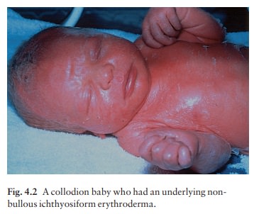
This
is a description and not a diagnosis. The bizarre skin charges are seen at
birth. At first the stratum corneum is smooth and shiny, and the skin looks as
though it has been covered with cellophane or col-lodion. Its tightness may
cause ectropion and feeding difficulties. The shiny outer surface is shed
within a few days leaving behind, most often, a non-bullous
Problems with temperature
regulation and high water loss through the skin in the early days of life are
best dealt with by the use of a high humidity incubator. Regular applications
of a greasy emollient also limit fluid loss and make the skin supple. The much
rarer ŌĆśharlequin fetusŌĆÖ is covered with thick fissured hyperkeratosis.
Ectropion is extreme and most affected infants die early.
Lamellar ichthyosis and non-bullous ichthyosiform erythroderma
Understandably,
these rare conditions have often been confused in the past. Both may be
inherited as an autosomal recessive trait, and in both the skin changes at
birth are those of a collodion baby . Later the two conditions can be
distinguished by the finer scaling and more obvious redness of non-bullous
ichthyosiform erythroderma. Both last for life and are sufficiently disfiguring
for the long-term use of acitretin to be justifiable (Formulary 2). Lamellar
ichthyosis shows genetic heterogeneity: the most severe type is caused by
mutations in the gene for keratinocyte transglutaminase, an enzyme that cross-links
the cornified cell envelope, lying on chromosome 14q11.2.
Epidermolytic hyperkeratosis (bullous ichthyosiform erythroderma)
This
rare condition is inherited as an autosomal dominant disorder. Shortly after
birth the babyŌĆÖs skin becomes generally red and shows numerous blisters. The
redness fades over a few months, and the tend-ency to blister also lessens, but
during childhood a gross brownish warty hyperkeratosis appears, sometimes in a
roughly linear form and usually worst in the flexures. The histology is
distinctive: a thickened granular cell layer contains large granules, and
clefts may be seen in the upper epidermis. The condition is caused by mutations
in the genes (on chromosomes 12q13 and 17q21) controlling the production of
keratins 1 and 10. A few patients with localized areas of hyperkeratosis with
the same his-tological features have gonadal mosaicism, and so their children
are at risk of developing the general-ized form of the disorder. Treatment is
symptomatic and antibiotics may be needed if the blisters become infected.
Acitretin has helped in severe cases.
Other ichthyosiform disorders
Sometimes
ichthyotic skin changes are a minor part of a multisystem disease, but such
associations are very rare. RefsumŌĆÖs syndrome, an
autosomal recessive trait, is caused by deficiency of a single enzyme
con-cerned in the breakdown of phytanic acid, which then accumulates in the
tissues. The other features (retinal degeneration, peripheral neuropathy and
ataxia) over-shadow the minor dryness of the skin.
RudŌĆÖs
syndrome is an ichthyosiform erythrodermain association with mental
retardation and epilepsy. In NethertonŌĆÖs syndrome, brittle
hairs, with a so-calledŌĆśbamboo deformityŌĆÖ, are present as well as a curious
gyrate and erythematous hyperkeratotic eruption (ichthyosis linearis
circumflexa). Other conditions are identified by confusing acronyms: IBIDS
(also known as trichothiodystrophy) stands for Ichthyosis, Brittle
hair, Impaired intelligence, Decreased fertility and Short stature; the KID syndrome consists
of Keratitis, Ichthyosis and Deafness.
Acquired ichthyosis
It
is unusual for ichthyosis to appear for the first time in adult life; but if it
does, an underlying disease should be suspected. The most frequent is HodgkinŌĆÖs
disease. Other recorded causes include other lymphomas, leprosy, sarcoidosis,
malabsorption and a poor diet. The skin may also appear dry in hypothyroidism.
Related Topics