Chapter: Medical Physiology: Rhythmical Excitation of the Heart
The Sinus Node as the Pacemaker of the Heart
The Sinus Node as the Pacemaker of the Heart
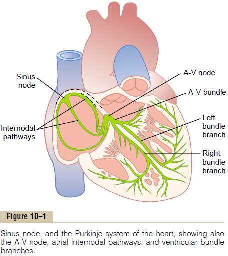
In the discussion thus far of the genesis and transmis-sion of the cardiac impulse through the heart, we have noted that the impulse normally arises in the sinus node. In some abnormal conditions, this is not the case. A few other parts of the heart can exhibit intrinsic rhythmical excitation in the same way that the sinus nodal fibers do; this is particularly true of the A-V nodal and Purkinje fibers.
The A-V nodal fibers, when not stimulated from some outside source, discharge at an intrinsic rhyth-mical rate of 40 to 60 times per minute, and the Purk-inje fibers discharge at a rate somewhere between 15 and 40 times per minute. These rates are in contrast to the normal rate of the sinus node of 70 to 80 times per minute.

The question we must ask is: Why does the sinus node rather than the A-V node or the Purkinje fibers control the heart’s rhythmicity? The answer derives from the fact that the discharge rate of the sinus node is considerably faster than the natural self-excitatory discharge rate of either the A-V node or the Purkinje fibers. Each time the sinus node discharges, its impulse is conducted into both the A-V node and the Purkinje fibers, also discharging their excitable membranes. But the sinus node discharges again before either the A-V node or the Purkinje fibers can reach their own thresh-olds for self-excitation. Therefore, the new impulse from the sinus node discharges both the A-V node and the Purkinje fibers before self-excitation can occur in either of these.
Thus, the sinus node controls the beat of the heart because its rate of rhythmical discharge is faster than that of any other part of the heart. Therefore, the sinus node is virtually always the pacemaker of the normal heart.
Abnormal Pacemakers—“Ectopic” Pacemaker. Occasionallysome other part of the heart develops a rhythmical dis-charge rate that is more rapid than that of the sinus node. For instance, this sometimes occurs in the A-V node or in the Purkinje fibers when one of these becomes abnormal. In either case, the pacemaker of the heart shifts from the sinus node to the A-V node or to the excited Purkinje fibers. Under rarer condi-tions, a place in the atrial or ventricular muscle devel-ops excessive excitability and becomes the pacemaker.
A pacemaker elsewhere than the sinus node is called an “ectopic” pacemaker. An ectopic pacemaker causes an abnormal sequence of contraction of the different parts of the heart and can cause significant debility of heart pumping.
Another cause of shift of the pacemaker is blockage of transmission of the cardiac impulse from the sinus node to the other parts of the heart. The new pace-maker then occurs most frequently at the A-V node or in the penetrating portion of the A-V bundle on the way to the ventricles.
When A-V block occurs—that is, when the cardiac impulse fails to pass from the atria into the ventricles through the A-V nodal and bundle system—the atria continue to beat at the normal rate of rhythm of the sinus node, while a new pacemaker usually develops in the Purkinje system of the ventricles and drives the ventricular muscle at a new rate somewhere between 15 and 40 beats per minute. After sudden A-V bundle block, the Purkinje system does not begin to emit its intrinsic rhythmical impulses until 5 to 20 seconds later because, before the blockage, the Purkinje fibers had been “overdriven” by the rapid sinus impulses and, consequently, are in a suppressed state. During these 5 to 20 seconds, the ventricles fail to pump blood, and the person faints after the first 4 to 5 seconds because of lack of blood flow to the brain. This delayed pickup of the heartbeat is called Stokes-Adams syndrome. If the delay period is too long, it can lead to death.
Related Topics