Chapter: Human Neuroanatomy(Fundamental and Clinical): Internal Structure of Brainstem
The Midbrain - Internal Structure of Brainstem
The Midbrain
Some features of the internal structure of the midbrain have been considered (Fig. 6.9). The subdivision of the midbrain into the tectum, the tegmentum, the substantia nigra, and the crus cerebri (or basis pedunculi) has been noted. The superior and inferior colliculi, the red nucleus and the reticular formation have been identified.
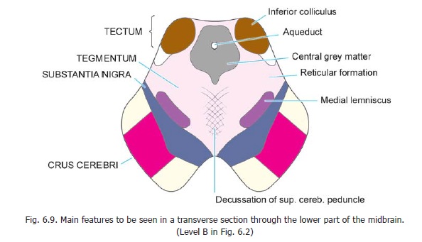
Transverse sections through the midbrain
A transverse section through the midbrain at the level of the inferior colliculus is shown in Fig. 11.8 and a section at the level of the superior colliculus in Fig. 11.9. We will first consider those features that are common to both these levels.
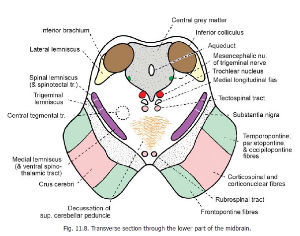
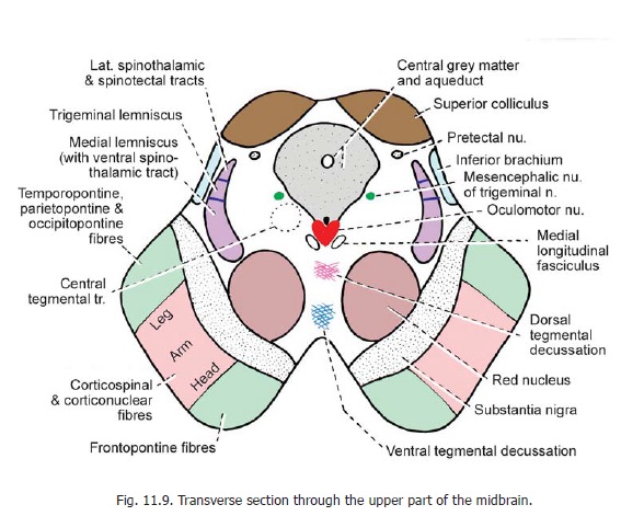
The crus cerebri (or basis pedunculi) consists of fibres descending from the cerebral cortex. Its medial one-sixth is occupied by corticopontine fibres descending from the frontal lobe; and the lateral one-sixth is occupied by similar fibres from the temporal, occipital and parietal lobes. The intermediate two-thirds of the crus cerebri are occupied by corticospinal and corticonuclear fibres. The fibres for the leg are most lateral and those for the head are most medial.
The substantia nigra lies immediately behind and medial to the basis pedunculi. It appears dark in unstained sections as neurons within it contain pigment (neuromelanin).
The substantia nigra is divisible into a dorsal part, the pars compacta; and a ventral part, the parsreticularis. The pars compacta contains dopaminergic and cholinergic neurons. Most of the neurons inthe pars reticularis are GABAergic. Superiorly, the pars reticularis becomes continuous with the globus pallidus. The substantia nigra is closely connected, functionally, with the corpus striatum (and in causation of Parkinsonism).
The midbrain is traversed by the cerebral aqueduct which is surrounded by central grey matter. Ventrally, the central grey matter is related to cranial nerve nuclei (oculomotor and trochlear). The region between the substantia nigra and the central grey matter is occupied by the reticular formation.
Section through midbrain at level of inferior colliculus
A section through the midbrain at the level of the inferior colliculus shows the following additional features (Fig. 11.8).
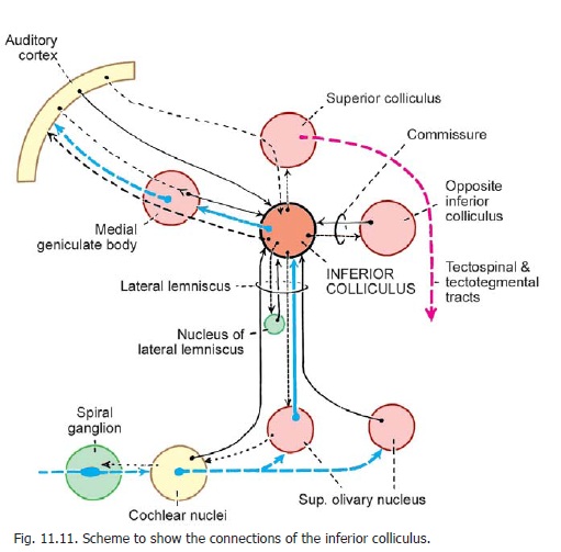
The inferior colliculus is a large mass of grey matter lying in the tectum. It forms a cell station in the auditory pathway and is probably concerned with reflexes involving auditory stimuli. Its connections are considered and are shown in Fig. 11.11.
The trochlear nucleus lies in the ventral part of the central grey matter. Fibres arising in this nucleus follow an unusual course. They run dorsally and decussate (in the superior medullary velum) before emerging on the dorsal aspect of the brainstem. The mesencephalic nucleus of thetrigeminal nerve lies in the lateral part of the central grey matter.
A compact bundle of fibres lies in the tegmentum dorsomedial to the substantia nigra. It consists of the medial lemniscus, the trigeminal lemniscus and the spinal lemniscus in that order from medial to lateral side. The medial lemniscus includes fibres of the ventral spinothalamic tract while the spinal lemniscus (made up mainly of the lateral spinothalamic tract) includes fibres of the spinotectal tract. More dorsally, the lateral lemniscus forms a bundle ventrolateral to the inferior colliculus (in which most of its fibres end). Important fibre bundles are also located near the middle line of the tegmentum. The medial longitudinal fasciculus lies ventral to the trochlear nucleus; and ventral to the fasciculus there is the tectospinal tract. The region ventral to the tectospinal tracts is occupied by decussating fibres of the superior cerebellar peduncle. These fibres have their origin in the dentate nucleus of the cerebellum. They cross the middle line in the lower part of the tegmentum. Some of these fibres end in the red nucleus while others ascend to the thalamus. The part of the tegmentum ventral to the decussation of the superior cerebellar peduncle is occupied by the rubrospinal tracts.
Section through midbrain at level of superior colliculus
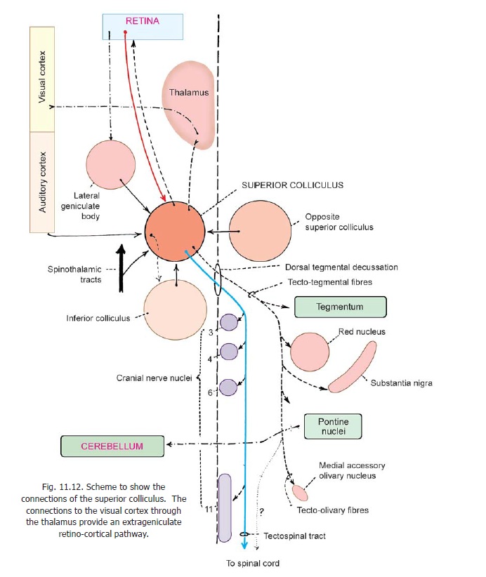
A section through the upper part of the midbrain (Fig. 11.9) shows two large masses of grey matter not seen at lower levels. These are the superior colliculus in the tectum, and the red nucleus in the tegmentum. The superior colliculus is a centre concerned with visual reflexes. Its connections are considered and are shown in Fig. 11.12.
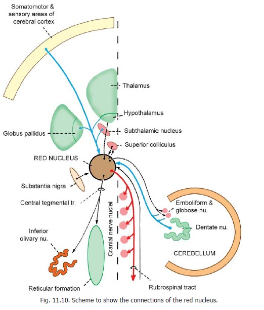
The red nucleus lies in the anterior part of the tegmentum dorsomedial to the substantia nigra. It is so called because of a reddish colour in fresh material. The colour is produced by the presence of an iron pigment in its neurons. The connections of the red nucleus are considered below and are shown in Fig. 11.10.
The oculomotor nucleus lies in relation to the ventral part of the central grey matter. The nuclei of the two sides lie close together forming a single complex. TheEdinger Westphal nucleus (which supplies the sphincter pupillae and ciliaris muscle) forms part of the oculomotor complex. The oculomotor complex is related ventrally to the medial longitudinal fasciculus.
Closely related to the cranial part of the superior colliculus there is a small collection of neurons that constitute the pretectal nucleus. This nucleus is concerned with the pathway for the pupillary light reflex.
The pretectal nucleus extends cranially to the junction of the midbrain with the diencephalon. It receives retinal fibres through the optic tract. It also receives some fibres from the superior colliculus and from the visual cortex. The main efferents of the nucleus reach the oculomotor nuclei (of both sides). Some efferents reach the superior colliculus and the pulvinar.
The bundle of ascending fibres consisting of the medial lemniscus, the trigeminal lemniscus and the spinal lemniscus lies more dorsally than at lower levels (because of the presence of the red nucleus). The lateral lemniscus is not seen at this level as its fibres end in the inferior colliculus. However, the inferior brachium that conveys auditory fibres to the medial geniculate body can be seen near the surface of the teg-mentum. The region of the tegmentum near the middle line shows two groups of decussating fibres.
The dorsal tegmental decussation consists of fibres that have their origin in the superior colliculus and cross to the opposite side to descend as the tectospinal tract. The ventral tegmental decussation consists of fibres that originate in the red nucleus and decussate to form the rubrospinal tracts.
Connections of the red nucleus
The red nucleus consists of a cranial parvicellular part and a caudal magnocellular part. The magnocellular part is prominent in lower species, but in man it is much reduced and is distinctly smaller than the parvicellular part.
The red nucleus receives its main afferents from:
a. the cerebral cortex (directly, and as collaterals from the corticospinal tract); and
b. the cerebellum (dentate, emboliform and globose nuclei).
The efferents of the nucleus are as follows. The magnocellular part projects to the spinal cord through the rubrospinal tract; and to motor cranial nerve nuclei (III, IV, V, VI, VII) through the rubrobulbar tract. Some fibres reach the reticular formation. The parvicellular part of the nucleus gives origin to fibres that descend through thecentral tegmental fasciculus to reach the inferior olivary nucleus. Some fibres reach the reticular formation. Other connections of the red nucleus are shown in Fig. 11.10
Inferior Colliculus
The inferior colliculus is an important relay centre in the acoustic (auditory) pathway. It receives fibres of the lateral lemniscus arising in the superior olivary complex. Each colliculus receives auditory impulses from both ears. These impulses are relayed to the medial geniculate body (the fibres passing through the inferior brachium) and from there to the acoustic (auditory) area of the cerebral cortex.
Other connections of the inferior colliculus are shown in Fig. 11.11 which illustrates the complete auditory pathway. Note that the inferior colliculus can influence motor neurons in the spinal cord and brainstem through the superior colliculus and the tectospinal and tectotegmental tracts.
Traditionally, the inferior colliculi have been regarded as reflex centres for responses to auditory stimuli. The colliculi are important in differentiating sounds received by the two ears, and thus, in locating the source of sound.
Each inferior colliculus has a main nucleus (placed centrally). This nucleus is divisible into dorsomedial and ventrolateral zones. More superficially, in the colliculus, there is a dorsal cortex which is divided into four laminae. The neurons of the inferior colliculus are arranged in groups responding todifferent frequencies of sound. Those in the dorsal part of the central nucleus respond to low frequencies, while ventrally placed cells respond to higher frequencies. Recent studies suggest that a similar arrangement probably exists at all levels of the auditory pathway.
Lesions of the inferior colliculus produce defects in appreciation of tones, of localisation of sound, and of reflex movements in response to sound.
In Fig. 11.11 note that the various auditory centres are also interconnected by fibres running in a direction opposite to that of the main auditory pathway. Thesedescending auditory fibres probably have a regulatory role on conduction through the pathway.
Connections of the Superior Colliculus
The superior colliculus has a complex laminar structure, being made up of seven layers. Its connections are shown in Fig. 11.12. Its most important afferents are those that bring visual impulses from the retina. The major efferents are the tectospinal tract, and tectonuclear fibres to the nuclei of cranial nerves responsible for moving the eyes and head. Some efferents also reach the retina. The colliculus has, therefore, been regarded as a centre for reflex movements of the head and eyes in response to visual stimuli.
However, recent work has shown that the functions of the superior colliculi may be much more complex. In addition to visual impulses the superior colliculi receive auditory impulses (through the inferior colliculi), and somatic impulses (touch, pain, temperature) through collaterals of spinothalamic tracts. They also receive fibres from the temporal and occipital cerebral cortex. The colliculi send efferents to various centres in the brainstem (red nucleus, substantia nigra, reticular formation) and can influence the cerebellum through the pontine nuclei. The fibres to the reticular formation reach reticular nuclei in the midbrain, pons and medulla
From these connections it appears likely that the superior colliculi are concerned with complex interactions between visual inputs and various activities of the body. Some fibres descend to the superior colliculus from the auditory cortex, and may be involved in integration of visual and auditory behaviour.
Connections of the Substantia Nigra
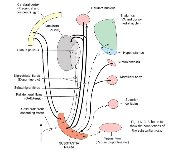
The main connections (both afferent and efferent) are with the striatum (i.e., caudate nucleus and putamen). Dopamine produced by neurons in the substantia nigra (pars compacta) passes along their axons to the striatum (mesostriatal dopamine system). Dopamine is much reduced in patients with a disease called Parkinsonism in which there is a degeneration of the striatum.
Other connections of the substantia nigra are shown in Figs. 11.13, 11.14. The functional relationships of the substantia nigra with the basal nuclei are considered..

Along with other groups of dopaminergic neurons present in the ventral part of the tegmentum, the substantia nigra is believed to be a neural centre for “adaptive behaviour”. Efferents of this system are widely distributed.
Some fibre bundles seen in the brainstem
In addition to the various ascending and descending tracts described later, there are a number of fibre bundles to be seen in the brainstem. These include:
1. The medial longitudinal fasciculus (or bundle)
2. The central tegmental tract.
3. The dorsal longitudinal fasciculus.
Parts of the medial forebrain bundle, and of the mamillary peduncle are also seen.
Related Topics