Chapter: Human Neuroanatomy(Fundamental and Clinical): Internal Structure of Brainstem
Control of Eye Movements - Internal Structure of Brainstem
Control of Eye Movements
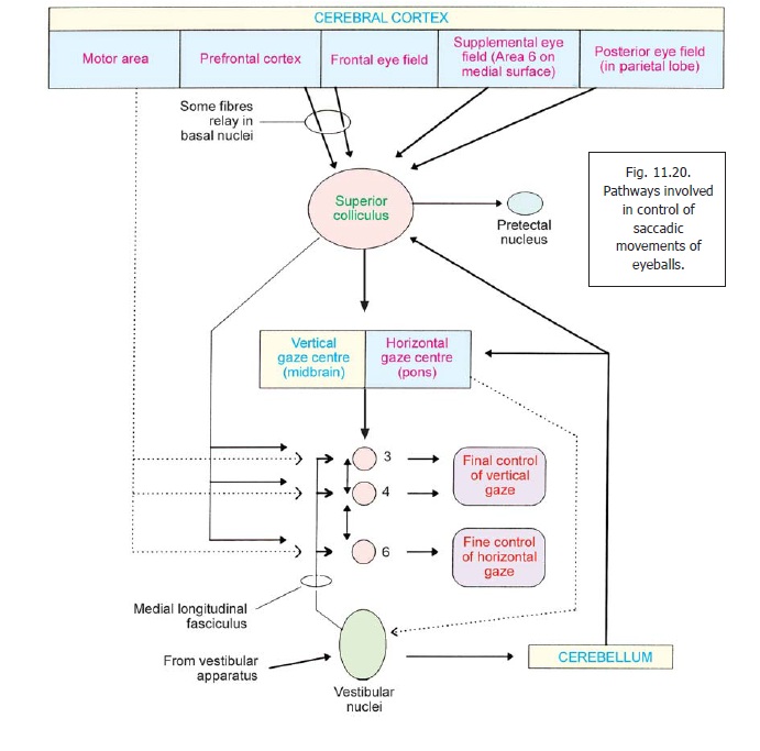
The cranial nerve nuclei that innervate the muscles that move the eyeballs are under the influence of a complex network involving the cerebral cortex, the cerebellum, the superior colliculus, the vestibular nuclei and other centres (Figs. 11.20, 11.21). As many of the centres involved lie in the brainstem it is convenient to consider the entire system here.
The gross control of ocular movements is similar to that for other movements. The motor area of the cerebral cortex projects to the cranial nerve nuclei concerned through corticonuclear fibres. The nuclei are also influenced by other centres. In this section we will consider some aspects of the control of fine movements of the eyeballs.
When the gaze is shifted from one object to another the eyes undergo sharp movements called saccades. The controlling centres determine the velocity and extent of a saccade. A different kind of control is required when the eyes follow a moving object (pursuit movement).
(a) The areas of cerebral cortex involved in programming and co-ordination of all eye movements are:
a. the frontal eye field (area 8);
b. the supplemental eye field located on the medial surface of the hemisphere (area 6).
c. the prefrontal cortex (anterior to the frontal eye field); and
d. the posterior eye field (in the parietal lobe).
(b) The areas of cerebral cortex listed above project to the superior colliculus (some of them passing via the basal nuclei). The superior colliculus also receives fibres from the retinae, from pretectal nuclei, and from the cerebellum.
(c) The superior colliculus projects to two important centres which are as follows.
1) Thevertical gaze centrelies in the midbrain. It is located in the rostral interstitial nucleus (acollection of neurons within the medial longitudinal fasciculus), and in the interstitial nucleus of Cajal. This centre controls vertical eye movements.
2) Thehorizontal gaze centrelies in the pons, in the paramedian reticular formation. Thecentre controls horizontal eye movements.
In addition to the afferents from the superior colliculus, these centres also receive:
a. some direct cortical fibres; and
b. fibres from the cerebellum.
Efferents from the vertical and horizontal gaze centres project to the oculomotor, trochlear and abducent nuclei.
(d) The cranial nerve nuclei supplying muscles of the eyeball receive the following afferents.
a. Corticonuclear fibres.
b. Fibres from the vertical and horizontal gaze centres described above.
c. Direct fibres from the superior colliculus and from pretectal nuclei.
d. Fibres from vestibular nuclei. These travel through the medial longitudinal fasciculus. They provide information about movements of the head and neck (from the vestibular apparatus). Through the vestibular nuclei, the oculomotor trochlear and abducent nuclei are brought under cerebellar control.
(e) The final control of vertical gaze is by fibres passing from the oculomotor and trochlear nuclei (to reach muscles producing upward and downward movements of the eyeballs). The final control of horizontal gaze is mainly by the abducent nerve (lateral rectus), but is also influenced by the oculomotor nerve (medial rectus).
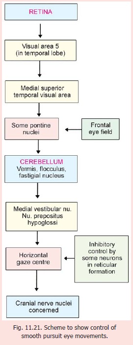
The horizontal gaze centre is also involved in control of eye movements when the gaze follows a moving object. These are called ‘pursuit movements’. The control of these movements is complex and is summarised in Fig. 11.21.
Injury to the medulla is usually fatal because vital centres controlling the heart and respiration are located here. As tracts are closely packed as they pass through the brainstem lesions produce widespread effect. Paralysis due to a lesion in the medulla is calledbulbar palsy. In this condition the 9th, 10th, 11th and 12th cranial nerves are affected.
Damage to corticospinal tracts in the brainstem roduces various syndromes. See Weber’s syndrome,Raymond’s syndrome andMillard Gubler syndrome.
Abnormality in upward gaze can occur in tumours of the pineal body. This is called Parinaud’ssyndrome.
Related Topics