Chapter: Biology: Practical Botany
Study of the Structure of Plant and Animal Cells

STUDY OF THE STRUCTURE OF PLANT
AND ANIMAL CELLS:
Requirements: Compound microscope, slide, cover slip, forceps, water, onion.
Procedure-1: Take a fleshy scale leaf from a fresh onion and pick up a thinmembrane
from its outer epidermis with the help of a forceps. Place the thin membrane on
the slide in a drop of water. Cover the piece with a cover slip and observe
under microscope.
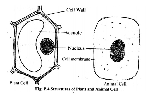
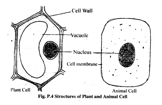
Observation: Under high power objective a single layer of cells will be seen.The
cells are with a definite shape and each of them is surrounded by a thick cell
wall. Near the cell wall, a nucleus is presenting in the cytoplasm. In every
cell there is a large vacuole.

Procedure-2: Wash your hands properly and bring out a thin membrane frominside your
mouth rubbing with your finger. Now rub the membrane, on your finger, over the
slide. A sticky layer will form on the slide. Then observe the sticky area of
the slide under a compound microscope.
Observation: Cell will be seen under the microscope. The cells are more orless round
in shape, a thin membrane surrounds each cell but there is no cell wall. The
nucleus is present at centre of the cell and there is no vacuole.

Related Topics