Chapter: 11th Microbiology : Chapter 7 : Morphology of Bacteria
Structures External to Cell Wall of Bacetria
Structures External to Cell Wall
of Bacetria
Appendages
Flagella
Flagella (singular flagellum) are threadlike, long, thin helical
filaments measuring 0.01-0.02nm in diameter. These appendages extend outward
from the plasma membrane and cell wall. Flagella are so thin that they cannot
be observed directly with a bright field microscope, but must be stained with
special techniques (example: Fontana’s silver staining technique) that increase
their thickness. The detailed structure of a flagellum can only be seen in the
electron microscope.
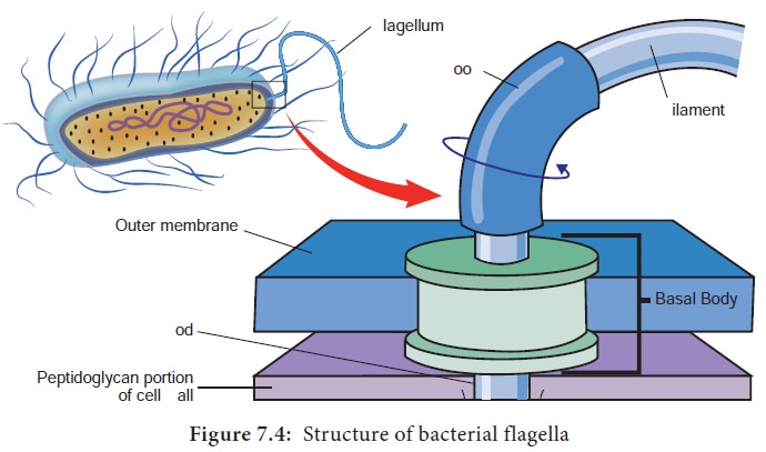
The bacterial flagellum is composed of three parts: a basal body
(associated with the cytoplasmic membrane and cell wall), a short hook and a
helical filament (which is usually several times as long as the cell). Filament
is external to cell wall and is connected to the hook at cell surface; the hook
and basal body are embedded in the cell envelope (Figure 7.4). Hook and
filament are composed of protein subunits called as flagellin.
One can generalize that all spirilla, about half of the bacilli
and a small number of cocci are flagellated. Some bacteria do not have
flagella. Flagella vary both in number and arrangement on the cell surface.
Flagella are arranged generally in two patterns.
1. In polar arrangement, the flagella are attached at one or
both ends of the cell. Bacteria with polar flagellar arrangement are further
classified into monotrichous, lophotrichous, and amphitrichous.
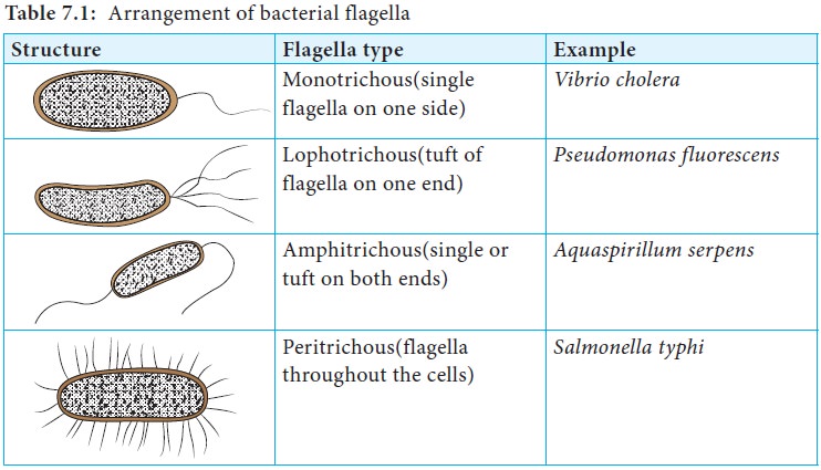
2. In lateral arrangement, flagella are arranged randomly all
over the surface of the cell. Bacteria with lateral flagellar arrangement are
called peritrichous. (Table 7.1)
Various types of mobility are observed based on the arrangement
of the flagella. Serpentine motility is seen with Salmonella, darting motility with Vibrio and tumbling motility with Listeria monocytogenes.
Some bacteria like Cytophaga exhibit
a gliding motility, which is slow sinuous flexing motion. This occurs when the
cells come in contact with solid surface.
Some bacteria have the ability to move toward or away from chemical substance. This movement is called chemotaxis. Positive chemotaxis is the movement of a cell in the direction of a favorable chemical stimulus (usually a nutrient). Negative chemotaxis is the movement away from a chemical substance (usually harmful compound). Some photosynthetic bacteria exhibit phototaxis, movement in response to light rather than chemicals.
The presence of motility is one piece of information used to
identify a pathogen in the laboratory. One way to detect motility is to stab a
tiny mass of cells into soft (semi solid) medium in a test tube. Growth
spreading rapidly through the entire medium is indicative of motility.
Alternatively, cells can be observed microscopically by a hanging drop method.
Pili
Pili (singular pilus) are straight, short and thin and more
numerous than flagella around the cell. They can be observed only by electron
microscopy. They are found only in certain species of Gram negative bacteria.
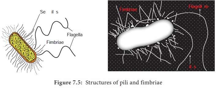
Pili play no role in motility. Pili originate from the plasma membrane and are
made up of a special protein called pilin (Figure 7.5).
Pili play a major role in human infection by allowing pathogenic
bacteria to attach to epithelial cells lining the respiratory, intestinal or
genitourinary tracts. This attachment prevents the bacteria being washed away
by body fluids, thus helps in establishment of infection. One specialized type
of pilus (sex pilus) helps in the transfer of genetic material between the
bacterial cells. This process is called conjugation.
Fimbriae
Fimbriae (singular: fimbria) is another term used for short pili that occur in great number around the cell. They enable bacteria to attach to surfaces and to each other, so that the bacteria form clumps or films called pellicles on the surface of liquid in which they are growing. Fimbriae are found in Gram positive as well as in Gram negative bacteria.
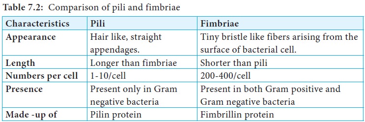
Table 7.2 compares the pili and fimbriae.
Extracellular Polymeric Substance (EPS)
Many bacteria secrete high molecular weight polymers that adhere
to the exterior of the cell wall to form a capsule or slime layer. Glycocalyx
is often used to refer to any polysaccharide material outside the cell wall.
Capsules and slime layer are considered to be glycocalyxes (Table 7.3).
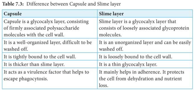
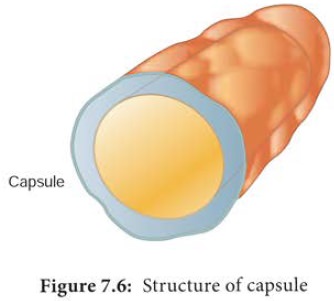
Capsules
Some bacterial cells are surrounded by a viscous substance
forming a covering layer or envelope around the cell wall called capsule
(Figure 7.6). Capsule is usually made up of polysaccharide.
It may be homopolysaccharide (made up of a single kind of sugar) or heteropolysaccharide (made up of several kinds of sugars). These are synthesized from sugars within the cell, transported and polymerized outside the cell. The capsule of some bacteria is made of polypeptides. The capsule of Bacillus anthracis has polymer of D-glutamic acid. Capsules are highly impermeable. Capsules can be demonstrated using special staining technique utilizing Indian ink or with Nigrosin stain. The presence of capsule in fresh isolates gives a moist and shiny appearance to the bacterial colonies on an agar medium. Capsular material is antigenic and may be demonstrated by serological methods.
The role of the capsule varies depending on the bacterium.
A thick capsule protects cells from dehydration.
Capsules protect the pathogenic bacteria from being engulfed and
destroyed by white blood cells (phagocytes).
Capsules are virulence factors of many pathogenic bacteria, such as Streptococus pneumonia, Haemophilius influenza and Bacillus anthracis. Encapsulated bacterial cells generally have greater virulence.
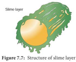
Slime layer
Some bacteria are covered with a surface layer that is loosely
distributed around the cell and diffuses into the medium, this surface layer is
referred to as slime layer. (Figure 7.7) The slime layer is a structure that is
easily washed off. Slime layer protects bacteria from loss of water and
nutrients. Slime has little affinity for basic dyes and is ivisible in Gram
stained smears.
Other Appendages
Sheath
Sheathed bacteria are bacteria that grow as long filaments in
the form of chain or trichome. These bacteria are enclosed by a hollow tube
like structure known as sheath
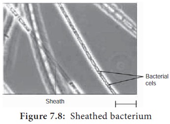
Within the shealth, the bacteria are capable of growth and division. Aquatic bacteria mostly form sheath. Examples of sheathed bacteria include Leptothrix discophora (also known as iron bacteria), Sphaerotilus and Clonothrix.
Function:
·
It provides mechanical support.
·
In a few bacteria, shealth is strengthened by the deposition of
ferric and manganese hydroxides.
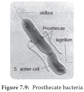
Prosthecae
They are semi rigid extensions of cell wall and cell membrane.
Some bacteria may contain more than one prosthecae (Figure 7.9). Aerobic
bacteria in fresh water and marine environment possess prosthecae. Some of the
prosthecate bacteria are Caulobacter,
Stellar, Prosthecobacter and
Hyphomicrobium.
Function:
·
Prosthecae increase surface area for absorption of nutrients
from the dilute aquatic environment.
·
Helps in adhesion.
·
Some prosthecae develop bud at the tip and helps in asexual
reproduction.
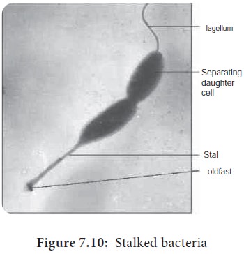
Stalk
It is a nonliving ribbon like tubular structure. It is formed by
excretory product of bacteria. Some of the stalked bacteria are Gallionella, Planctomyces (Figure 7.10).
Function:
Stalk helps in attachment of cells to solid surface.
Related Topics