Practical Experiment | Microbiology - Spotters: Slide | 12th Microbiology : Practical Experiment Manual
Chapter: 12th Microbiology : Practical Experiment Manual
Spotters: Slide
B) Slide
6. Cyst of Entamoeba histolytica
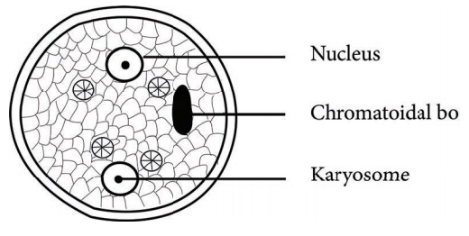
• Cyst is
one of the three forms of entamoeba histolytica
• A
mature cyst is a quadrinucleate spherical bod
• Mature
cysts are passed in the stool of infected person
• Direct
examination of wet mount of stool for cysts is diagnostic of intestinal
amoebiasis
7. Penicillium species
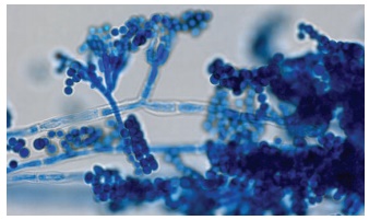
• Colony
of penicillium are initially white and fluffy and later produce pigmented
spores and turn into shades of green or blue green
• Hyphae
are hyaline and septate
• Condiophores
are long, give rise to branching phialids
• Phialids
branch and give the appearance of brush or penicillins
• They produce sterigmata bearing chain of conidia (spores) which are oval or spherical and measure 1-2micrometer.
8. Microfilariae
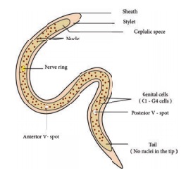
• Filariasis
is caused by nematodes (roundworms) like Wuchereria bancrofti that inhabit the
lymphatics and subcutaneous tissues.
• The
female worms release the firsstage larvae called microfilariae, which are
detected in the peripheral blood.
• Identification
of microfilariae by microscopic examination is the most practical diagnostic
procedure.
• The
blood sample can be a thick smear, stained with Giemsa.
• The larva measures about 290microns in length and 6-7micron in breath.
9. Egg of Ascaris lumbricoides
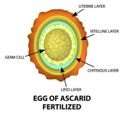
• These are passed in stool of the infected host.
• Brownish due to bile pigment.
• Fertilised eggs are rounded and have a thick
shell (chitinous).
• Unfertilised eggs are elongated and larger than fertile
eggs.
• When
ingested through water or contaminated food by human it causes Ascariasis.
• Microscopic identification of eggs in the stool
is the most common method for diagnosing intestinal ascariasis.
10. Heterocysts of Nostoc
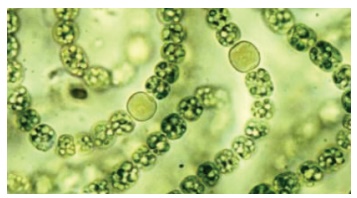
• Heterocysts are specialized structures having
thick cell wall formed in some filamentous blue green algae like Nostoc,
Anabena.
• They may be terminal or found in between the
vegetative cells attached to it by means of pores.
• They are sites of atmospheric nitrogen fixation.
• They serve as a store house of food material.
11. Acid
fast bacilli
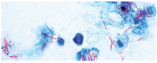
• Acid fast bacilli contains mycolic acid in their
cell walls hence do not get stained easily, however once stained cannot be
decolourised easily.
• Special method like Ziehl- Neelson’s Carbol
fuchsin is used to stain acid fast bacilli.
• The acid- fast bacilli are stained red in colour
while the non acid fast cells appear blue when counterstained with methylene
blue.
• Mycobacterium tuberculosis is and acid fast bacilli.
Related Topics