Practical Experiment | Microbiology - Blood Staining | 12th Microbiology : Practical Experiment Manual
Chapter: 12th Microbiology : Practical Experiment Manual
Blood Staining
Blood Staining
AIM
To make a
blood smear ,stain it using Field’s stain and observe the erythrocytes and
leucocytes.
Theory and Principle:
Blood
smears are used to determine leukocyte differentials, to evaluate erythrocyte,
platelet and leukocyte morphology, and, if necessary, to estimate platelet and
leukocyte counts. It is also used for diagnosis of parasites like plasmodium in
the blood.
Field’s
Stain is a romanowsky stain, used for rapid processing of blood specimens and
is used to stain thick and thin films. It consists of two differential stain.Field stain A which is methylene blue
and Azure dissolved in a phosphate buffer solution.It is the basic component of
the stain and Field stain B made up
of Eosin Y in a buffer solution which is the acidic component of the
stain.These basic and acidic dyes induce several colours when applied to
cells.The fixator, methanol, does not allow any additional changes to the
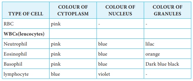
slide. The basic component of peripheral white blood cell( cytoplasm) is
stained with acid dye and the acid component that is nucleic acid of the
nucleus takes on the basic dye and is stained blue to violet. The neutral components
of the cells are stained by both dyes(Field’s stain A and B solution).
Requirements
• Cotton
• Spirit
• Blood
sample
• Clean
grease free slides
• Methanol
fixative
• Field’s
stain A and Field’s stain B.
Procedure
1. Finger Prick under aseptic condition.
2. Place a small drop of blood, on one side about
1-2 cm from one end of a slide.
3. Without delay place another slide at an angle of
45° to make contact with the drop.
4. Spread it over an area of about 2 cm2(The film
should be distributed so thinly that it appears transparent.
5. After air drying the thin blood film,immerse or
fix the smear in methanol for 1 minutes.
6. Flood or
dip the slide in Field’s Stain A for 2-3 seconds.
7. Wash it with distilled water,
8. Flood or dip the slide in Field’s Stain B for
2-3 seconds and wash with distilled water.
9. Now air dry the smear and observe under
microscope
Diagram
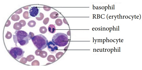
Observation
Results
The blood
smear was stained using field’s stain and erythrocytes and leucocytes were observed
under microscope.
Related Topics