Chapter: Genetics and Molecular Biology: Oncogenesis, Molecular Aspects
Recessive Oncogenic Mutations, Tumor Suppressors
Recessive Oncogenic Mutations, Tumor Suppressors
Transformation studies like those used to find the
DNA change associ-ated with a bladder cancer have shown that only about 10% of
tumors can be associated with dominant mutations. Similarly, fusion of
trans-formed tumor cells and normal cells shows that in most cases, the
genotype of the normal cells can suppress the oncogene being expressed in the
transformed cells. If the fusion cells possess an unstable karyotype and lose
chromosomes, then the descendants of these fused cells can again express the
transformed phenotype. Thus, oncogenesis can derive from two different
pathways. The first is dominant mutations that generate transformed cells
independent of the other cellular genes, and the second is a product of two
events. One of these is a mutation that would produce uncontrolled cell growth
were it not for the presence of another cellular regulator, and the second is
the loss of the regulator.
The simplest example of the two-step pathway is a
mutation in a growth factor such that it no longer prevents uncontrolled
growth. Ordinarily the mutation to inability to repress growth is not revealed
because the mutation is recessive and the nonmutated copy of the gene product
from the other copy of the chromosome suppresses growth. If, however, the
nonmutated copy is lost by recombination or mutated, then the growth inhibition
ceases. Retinoblastoma is a form of cancer that develops in young children.
Most cases of retinoblastoma are familial, and the pattern of inheritance
indicates that the gene involved is not on a sex chromosome. The patterns of
retinoblastoma develop-ment suggest that in addition to inheriting a defective
gene, a second event must occur in the stricken individuals. We now know that
this second event frequently is recombination of the defect from one
chro-mosome onto the homologous chromosome in a way that leaves both
chromosomes defective for the critical gene. Gene conversion as dis-cussed
earlier could do this.
Through good luck and hard work the gene whose
defect gives rise to retinoblastoma was cloned. The first step in the process
came from the finding that an appreciable fraction of retinoblastoma cell lines
possess a gross defect or deletion in part of chromosome 13. This suggests that
the gene in question lies on chromosome 13. With this as a starting point it
was possible to clone the gene. The original idea was that perhaps a clone
could be found containing some of the DNA that is deleted from chromosome 13 in
some of the retinoblastoma lines. By chromosome walking, it might then be
possible to clone the desired retinoblastoma gene.
A library of about 1,000 clones of segments of
chromosome 13 was available. These were then screened against a large panel of
retinoblas - toma lines to see if any of the clones contained DNA absent from
the transformed cell lines. One clone indeed contained DNA absent from two of
the retinoblastoma lines. Best of all, it was complementary to an RNA that is
synthesized in normal cells, but not in retinoblastoma cells. This suggests
that the protein could, in fact, be the gene for the suppres-sor of
retinoblastoma.

Further study of the retinoblastoma protein
required sensitive assays for its presence. Two approaches were used to make
antibodies against the supposed retinoblastoma suppressor protein. Part of the
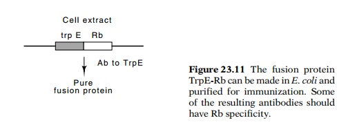
cloned and sequenced gene was fused to the trpE
gene and a fusion protein was synthesized in E. coli, purified by
making use of the trp tag, and used
to immunize animals for antibody synthesis (Fig. 23.11). The second approach
was to synthesize chemically peptides corresponding to sev-eral regions of the
presumed protein and use these to immunize ani-mals.
With antibodies against the protein, the synthesis
and properties of the retinoblastoma protein could be studied. It is indeed
synthesized in normal cells, but frequently is missing in retinoblastoma cells.
The protein is phosphorylated, and has a molecular weight of about 105 kD. This
is the molecular weight of one of the cellular proteins to which the adenovirus
E1a protein, the papillomavirus E7 protein, and the SV40 T antigen bind. Since
the E1a-associated protein is phosphorylated like the Rb protein, and since
phosphorylation of proteins is relatively rare, it seemed possible that the
retinoblastoma suppressor protein and the E1a-associated protein were one and
the same. Indeed, they were. Both antibody specificity and proteolysis patterns
confirm the identity. Thus it appears that the normal role of the
retinoblastoma suppressor gene is to counteract the positive activity of some
other cell growth inducing pathway. Hence one of the steps in transformation by
adenovirus, papillomavirus, and SV40 is to inactivate suppressor proteins
rather than to synthesize a dominant growth-inducing gene in the cells.
In as many as 50% of human tumors, the p53 protein
is found to have been altered. Thus, this protein appears to play a key role in
regulating cell growth. What is the nature of the regulation expressed by the
p53 protein? Is it required at all times for proper cell growth or regulation
of differentiation? A conceptually simple way to answer these questions is to
attempt to make a homozygous mouse deficient in p53 protein. Such mice turn out
to develop normally, but they are highly likely to develop cancers before they
are as old as six months. The tumors are found in many different tissues.
Further study has shown that p53 prevents the onset of DNA replication in a
cell possessing damaged DNA or a broken chromosome. By blocking DNA synthesis,
and allowing time for DNA repair, the spontaneous mutation frequency in cells
is greatly reduced.
Related Topics