Chapter: 11th 12th std standard Class Nursing Health Care Hospital Hygiene Higher secondary school College Notes
Physiology of Human Reproduction
Physiology of reproduction:
Menstrual cycle or uterine cycle:
It
is a series of changes in the uterus resulting in the discharge of blood from
the vagina each month. Menstruation can be defined as, 'sloughing and discharge
of the lining of the uterus if conception does not take place.' This time
varies in different women and also from time to time-in same woman.
The first day of the cycle is the
first day when bleeding begins. The ovarian hormones control the menstrual
cycle. There are three main phases and they affect the tissue structures of the
endometrium. The average time of menstrual cycle is 28 days and recurs
regularly from puberty to menopause except in pregnancy. The three phases are:
1. Proliferative
phase: Follicular stimulating hormonal
level increases in blood,
stimulating oestrogen secretion, which causes the endometrium to thicken and
become more vascular. This phase follows menstruation and lasts until
ovulation.
2. Secretary
phase: The secretary phase follows ovulation
and is under the influence of
progesterone and oestrogen from the corpus luteum. Leutinising hormone level
increases in blood. Under the combined stimulus of estrogen and progesterone,
the endometrium reaches the peak of its thickening and vascularisation.
3. Menstrual
phase: It is characterised by vaginal
bleeding, lasts for 3 - 5 days. On
absence of fertilization, the thickened endometrium is shedded.
Two Gonadotrophic hormones are
released by the anterior pituitary gland. They are
1.
Follicular stimulating hormone:FSH is primarily responsible for stimulating the ovaries to secrete oestrogen and for
maturation of ovum.
2.
Luteinising Hormone (LH): ): LH is primarily responsible for stimulating the orpus luteum
for productoin of progesterone.
Puberty: This is the period in which, the reproductive organs develop and reach maturity. The first
signs are breast development and appearance of pubic hair. The body grows
considerably and takes on the female contour. Puberty culminates in the onset
of menstruation, the first period being called menarche. The first few cycles are not accompanied by ovulation.
Puberty usually occurs between 12 and 14 years.
Menopause: It is the end of a woman' s reproductive life, characterised by the gradual cessation
of menstruation. The period first becomes irregular and then ceases altogether.
This occurs between the ages of 45 to 50. It is the normal part of aging and
maturation. Menstruation ceases because the ovaries are no longer active. No
more ovarian hormones are produced. The reproductive organs become atrophied.
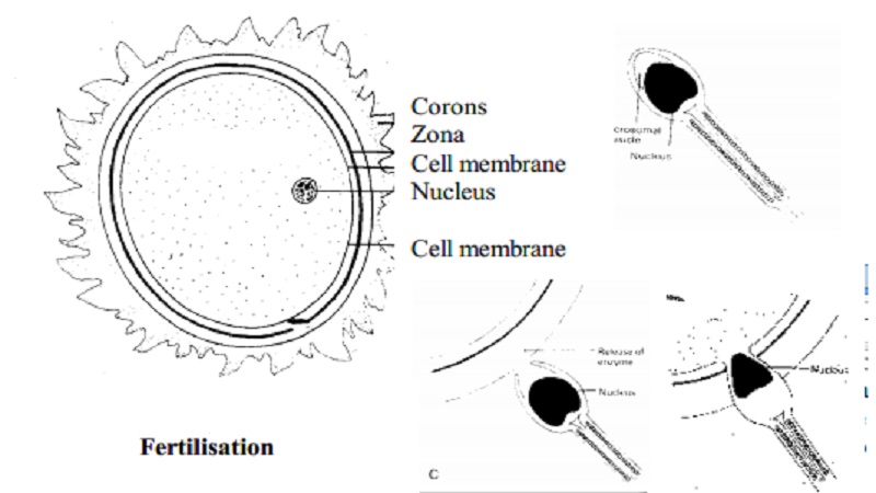
Fertilization: Following ovulation, the ovum about (0.15 mm) in diameter
passes into the fallopian tube and moves towards uterus. If coitus takes place
at this time, the alkaline mucus attracts the spermatozoa. About 300 million
sperms are deposited in the posterior fornix of the vagina. Those which are
propelled by the cervical mucus reach the fallopian tube and others are
destroyed by the acid medium of the vagina.
The matured sperm is capable of producing the enzyme
hyaluronidase, which allows the sperm to penetrate the cell membrane,
surrounding the ovum. Many sperm are needed for this, but only one will enter
into the ovum and fertilisation occurs. After this, the membrane is sealed to
prevent the entry of any further sperm and the nuclei of the two cells fuse.
The sperm and the ovum each
contribute half the complement of chromosomes to make a total of 46. The sperm
and ovum are known as the male and female gametes. The fertilised ovum is known
as the zygote.
Implantation of the fertilised ovum
(embedding) into the uterine cavity (endometrium) is called as nidation or
nesting. Normally this occurs by the 11th day after ovulation and
the endometrium closes over it completely.
Development of the fertilised ovum:
Fertilised ovum reaches the uterus by 3-4 days. Cell
division takes place as 2 into 4,8,16, etc, till a cluster of cells formed
known as morula (mulberry). Next a fluid filled cavity, a blastocele appears in
the morula and it is known as blastocyst. Outside of blastocyst there is a
single layer of cells known as trophoblast,
while the remaining cells are clumped together forming an inner cell mass.
The trophablast forms the placenta and chorion
while the inner cell mass become fetus
and amnion.
Formation of fetal membrane and
placenta:
The
trophoblast has two layers,
1.
Outer syncitiotrophoblast, which erodes the endometrium in the process of
embedding.
2.
The inner cytotrophoblast produces a hormone called human chorionic gonadotrophin (HCG) which reacts on corpus lutuem to continue the
pregnancy by producing oestrogen and progesterone.The trophoblast develops as
placenta which will nourish the fetus until delivery.
The
inner cell mass differentiates into three layers.
1.
From the ectoderm skin and nervous system are formed.
2.
From the mesoderm bones and muscles, heart and blood vessels and certain internal
organs are formed.
3.
From the endoderm mucous membranes and glands are formed.
During the first three weeks
following conceptual the fertilised ovum
is termed as zygote. From 3-8
weeks, it is termed as embryo. The organs and systems are
developed by 7th week. After 8 weeks, till birth it is termed as fetus.
Placenta:
The placenta is a remarkable organ. It originates from the
trophoblastic layer of the fertilised ovum. The placenta is completely formed
and starts functioning from 10 weeks after fertilisation.
The placenta is a round flat mass
about 20 cm in diameter and 2.5 cm thick at its center. It weighs approximately
1/6th of the baby' s weight. The fetus obtains oxygen and excretes
carbon dioxide through the placenta. Oxygen from the mother' s hemoglobin
passes into the fetal blood by diffusion. Similarly the fetus gives off carbon
dioxide into the maternal blood.
Functions of placenta:
The fetus obtains amino acids, glucose, vitamins, calcium,
phosphorus, iron and other minerals from the maternal blood through the
placenta.
1.
The placenta also stores glucose in
the form of glycogen. It also stores iron and fats soluble vitamins.
2.
The waste products such as carbon
dioxide, bilirubin and urea are excreted from the fetus through the placenta.
3.
The placenta prevents passing of
microorganisms from the mother to the fetus to some extent.
4.
The placenta also produces hormones
like the human chorionic gonadotrophic hormone, oestrogen, progesterone and
human placental lactogen (HPL).
The fetal sac:
The fetal sac consists of a double membrane, which contains
the fetus and the amniotic fluid.
1.
The chorion is the outer thick,
opaque membrane. It is derived from the trophoblastic cells.
2.
The amnion is the inner smooth tough
membrane. It is derived from the inner cell mass
Amniotic fluid (liquor amnii)
It is the clear, pale, straw-coloured fluid in which the
fetus floats. It is 500-800 ml in quantity at a term. Presence of amniotic
fluid allows for growth and free movement of the fetus and it acts as a shock
absorber thereby protecting the fetus.
Umbilical cord:
The cord extends from the fetus to the placenta and
transmits the umbilical blood vessels, two arteries and one vein. These are
enclosed by Wharton' s jelly.
The length of the cord is about 50 cm.
Fetal development:
Summary
of fetal growth and development: 0-4 weeks after conception:
1.
Rapid Growth.
2.
Formation of embryonic plate.
3.
Primitive central nervous system
forms.
4.
Heart develops and begins to beat.
Limb buds form.
4 - 8 weeks:
1.Very rapid cell division occurs.
2.Head and facial features develop.
3.All major organs laid down in primitive form.
4.External genitalia present but sex is not distinguishable.
5.Early movements visible on ultrasound from 6 weeks.
8 -12 weeks:
Eyelids
fuse.
Kidneys
begin to function and the fetus passes urine from weeks.
1.Fetal circulation starts functioning.
2.Sucking and swallowing present.
3.Sex apparent.
4.Moves freely (not felt by mother)
12 - 16 weeks:
1.Rapid skeletal development - visible on x-ray.
2.Meconium present in gut.
3.Lanugo appears.
4.Nasal septum and palate fuse.
16 - 20 weeks:
1.Quickening - mother feels fetal movements.
2.Fetal heart heard on auscultation.
3.Vernix caseosa appears.
4.Finger-nails can be seen.
5.Skin cells begin to be renewed.
20 - 24 weeks:
1.Most organs become capable of functioning.
2.Periods of sleep and activity present.
3.Responds to sound.
4.Skin red and wrinkled
24 - 28 weeks:
1.
Survival may be expected if born.
2.
Eyelids reopen.
3.
Respiratory movement present.
28 - 32 weeks:
1.
Begins to store iron and fat.
2.
Testes descend into scrotum.
3.
Lanugo disappears from face.
4.
Skin becomes pale and less wrinkled.
32 - 36 weeks:
1.
Increased fat makes the body more
rounded.
2.
Lanugo disappears from body.
3.
Head hair lengthens.
4.
Nails reach tips of fingers.
5.
Ear cartilage soft.
36 - 40
weeks:
1.
Term is reached and birth is due.
2.
Contours rounded.
3.
Skull firm.
Related Topics