Chapter: Genetics and Molecular Biology: Lambda Phage Genes and Regulatory Circuitry
Physical Structure of Lambda
The Physical Structure of Lambda
The lambda phage that has been studied in the
laboratory possesses an isometric head of diameter 650 Ă… and a tail 1,500 Ă…
long and 170 Ă… wide. A tail fiber extends an additional 200 Ă… from the tail.
This tail fiber makes a specific contact with a porin protein found in the
outer membrane of the host cell, and the phage DNA is injected through the tail
into the cell. Apparently in an early isolation of lambda, investigators chose
a mutant form of the virus that lacked tail fibers. The natural form possesses
a number of tail fibers that attach the phage to something other than the
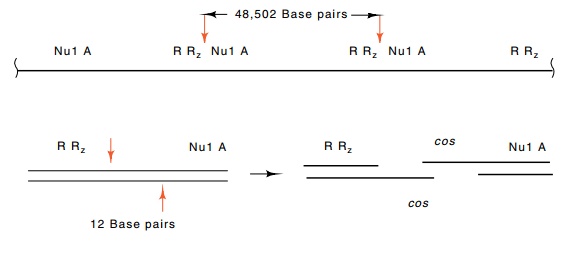
Figure
14.1 Cleavage at twocossites between theRandAgenes
generateslambda monomers from oligomers during encapsidation of lambda DNA.
Because each cos site is cut with a
12 base staggered cleavage, complementary end sequences are generated.

porin
protein. The DNA within the phage particle is double-stranded and linear, with
a length of 48,502 base pairs. This is roughly 1% the size of the Escherichia coli chromosome.
Intracellularly, lambda DNA exists as monomeric circles or polymeric circular
forms, but during encapsidation, unit-length lambda linear genomes are cut out
of the polymeric forms (Fig. 14.1). The cuts of the two DNA strands are offset
from one another by 12 bases. Thus the ends of the encapsidated phage DNA are
single-stranded and complementary.
Roughly
half the mass of a lambda particle is DNA and half is protein. Consequently,
the particle has a density in CsCl halfway between the density of DNA, 1.7
gm/cm3, and the density of protein, 1.3 gm/cm3. This
density facilitates purification of the phage since isopycnic banding in CsCl
density gradients easily separates the phage from most other cellular
components.
Related Topics