Chapter: Microbiology and Immunology: Bacteriology: Vibrio,Aeromonas,and Plesiomonas
Pathogenesis and Immunity - Vibrio cholerae
Pathogenesis and Immunity
Cholera is a toxin-mediated disease. Cholera toxin (CTx) pro-duced by V. cholerae is the key virulence factor of the bacteria.
◗ Virulence factors
Virulence factors of V. cholerae include (i) cholera toxin, (ii) toxin coregulated pilus (TCP), (iii) adhesin factor (ACF), (iv) hemagglutination-protease (hap; mucinase), (v) neuramini-dase, and (vi) siderophores (Table 35-4). Most of these virulence factors are expressed by multiple chromosomal genes present in V. cholerae. These genes include the genes for cholera toxin (CTx A and CTx B), TCP, ACF, hap, and neuraminidase.

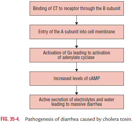
A1 fragment is responsible for the biological activity of the toxin. The A1 subunit stimulates cell-bound cyclic adenosine monophosphate (cAMP), which in turn converts ATP to cAMP in the intestines of epithelial cells of the gut. Cholera toxin has the following biological functions:
· This inhibits the absorption capacity and activates the excre-tory chloride transport in the intestinal enterocytes, even-tually leading to loss of sodium chloride in the intestinal lumen. The osmolality in the intestinal lumen is balanced by secretion of large quantities of water, which eventually overcomes the absorptive capacity and leads to diarrhea. The diarrheic fluid is isotonic in nature but contains much more of potassium and bicarbonate.
· It also inhibits absorption of sodium and chloride in the intestine.
· It also increases skin capillary permeability. Hence, it is also called as a permeability factor. The permeability factor can be detected by skin bluing test. In this test, cholera toxin is injected intradermally in rabbits or guinea pigs, followed by intravenous injection of Evans blue. In a positive test, the skin at the site of injection becomes blue due to increased capillary permeability.
The cholera toxin can be demonstrated by the following tests:
I. Rabbit ileal loop test was one of the classical methods developed by De and Chatterjee in 1952. It is a widely used method in which injection of V. cholerae culture or culture filtrate into the ligated ileal loop causes accumulation of fluid, leading to ballooning of the ileal loop.
II. ELISA and RIF (rapid immunofluorescence) tests.
III. Chemical estimation of cAMP in tumor cells that have been treated with the toxin.
IV. Demonstration of histological changes in adrenal tumor (Y1) cells, elongation of Chinese hamster ovary cells, and activation of lipolysis in rat testicular tissue.
Toxin coregulated pilus: The pili help in adherence ofV. cholerae to mucosal cells of the intestines.
Accessory colonization: These also help in adhesion of bacte-ria to the intestinal mucosa.
Hemagglutination-protease (mucinase): This enzyme, for-merly known as cholera lectin, is both agglutinin- and zinc-dependent protease. The enzyme splits mucus and fibronectin as well as subunit of the cholera toxin. It induces intestinal inflammation and also helps in releasing free vibrios from the bound mucosa to the intestinal lumen.
Neuraminidase: The enzyme destroys neuraminic acid,thereby increasing toxin receptors for the V. cholerae.
Siderophores: It is responsible for sequestration of iron.
◗ Pathogenesis of Cholera
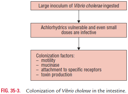
V. cholerae usually enters the body orally through contaminatedwater and food (Fig. 35-3). Gastric acidity is the first line of defense against infection caused by V. cholerae, because vibrios are highly susceptible to the acidity of stomach. The conditions that reduce acidity of the stomach to pH above 5 make the host more susceptible to infection by cholera vibrios.

On reaching the small intestine by using its own mecha-nisms, such as motility, chemokines, and production of enzymes (hemagglutinin and protease), V. cholerae reaches the mucous layer of the small intestine. Hemagglutinin and prote-ase break mucin and fibronectin of the mucosa. Subsequently, bacteria adhere to the intestinal wall facilitated by TCP. This synchronous action of CTx, TCP, and a few other virulence factors is regulated by Tox R gene products, which is known as the master switch.
Vibrios, once adhered to the intestinal wall, produce cholera toxin. The toxin activates cAMP, which inhibits the absorption of sodium transport and activates the excretory chloride trans-port in the intestinal epithelial cells. This leads to an accumu-lation of sodium chloride in the lumen of intestine. The high osmolarity of the intestinal fluid is balanced by large secre-tion of water, which overcomes the absorptive capacity of the lumen, eventually causing diarrhea (Fig. 35-4).

V. cholerae O139 strain shows a similar pathogenic mecha-nism to that of V. cholerae O1 except that it produces a unique O139 LPS and an immunologically related O antigen cap-sule. These two virulence factors enhance virulence of this organism. These are also responsible for increase in its resis-tance to human serum in vitro and development of bacte-remia occasionally seen during the infection caused by the bacteria.
◗ Host immunity
Immunity in cholera may be produced against the cholera toxin or against the bacteria. Natural infections in cholera induce some degree of immunity for a short duration of 6–12 months. Reinfection occurs in the same person after this period. Vaccination with killed cholera vaccine confers a short-lived protection. Live oral vaccines induce local immunity by pro-duction of IgA antibodies in the gut. The immunity induced by these oral vaccines is also short lived. An infection by V. cholerae biotype Classical usually protects against the infection caused by V. cholerae biotype Classical as well as Eltor strains. But infec-tion by V. cholerae biotype Eltor does not confer protection against biotype Classical.
Related Topics