Chapter: Microbiology and Immunology: Bacteriology: Neisseria
Pathogenesis and Immunity - Neisseria gonorrhoeae
Pathogenesis and Immunity
N. gonorrhoeae causes disease both by multiplying in tissues andby causing inflammation. The bacteria do not produce any toxins.
◗ Virulence factors
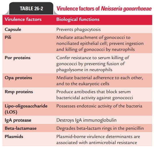
N. gonorrhoeae produces several virulence factors as mentionedbelow (Table 26-2):
Capsule: N. gonorrhoeaedoes not form a true carbohydratecapsule unlike N. meningitidis. Instead, it forms a polyphos-phate capsule, which is loosely associated with its cell surface. Capsule is most evident in freshly isolated gonococci and is antiphagocytic. It prevents phagocytosis of the gonococci.
Pili: Pili are hair-like structures that extend from the cyto-plasmic membrane through the outer membrane. The pili are composed of the proteins known as pilins, which are repeating

protein subunits. The expression of protein pilin is controlled by P gene complex. The pilins of all the strains of gonococci are antigenically different. There is a marked antigenic variation in gonococcal pili as a result of chromosomal rearrangement. More than 100 serotypes are known. The pili are important virulence factors:
· They play an important role in the virulence of the bacteria.
They mediate attachment of gonococci to nonciliated epi-thelial cells.
· They also contribute to virulence by preventing ingestion and killing of gonococci by neutrophils.
Other virulence factors: These include:
· Por protein of outer membrane protein (OMP) confers resis-tance to serum killing of gonococci by preventing fusion of phagolysosome in neutrophils.
· Opa proteins mediate bacterial adherence of bacteria to each other and to the eukaryotic cells.
· Rmp proteins produce antibodies that block serum bacteri-cidal activity against gonococci.
Lipo-oligosaccharide of the bacteria possesses endotoxic activity.
◗ Pathogenesis of gonorrhea
N. gonorrhoeae causes disease first by attaching themselves tomucosal cells. Subsequently, they enter the cells and multiply inside the cells and pass through the cells into the subepithe-lial space, thereby establishing the infection. Pili help in attach-ment of gonococci to mucosal surfaces and also contribute to the resistance by preventing ingestion and killing by PMN leukocytes. The outer membrane proteins, such as Opa proteins, facilitate adherence between gonococci and also increase adherence to phagocytes. The Opa proteins also facilitate sub-sequent migration of gonococci into the epithelial cells. The Por proteins inhibit phagolysosome fusion in the phagocytes, thereby protecting the phagocytosed bacteria from intracellu-lar killing. Production of beta-lactamase (penicillinase) by the bacteria also contributes to the invasion.
The host response is characterized by infiltration with leu-kocytes, followed by epithelial sloughing, formation of micro-abscesses in the submucosa, and production of purulent pus.
The LOS of gonococcal cell wall stimulates the production of tumor necrosis factor alpha (TNF-a) and other inflamma-tory responses which contribute to most of the symptoms asso-ciated with gonococcal infection.
◗ Host immunity
The main host defense mechanisms against gonococci are anti-bodies (IgA and IgG), complement, and neutrophils. Antibody response to gonococci is characterized by the production of serum IgG antibodies. IgG3 is the predominant immunoglobu-lin. Antibody response is strong against Opa proteins and LOS, whereas it is minimal against Por proteins. Antibodies to LOS cause activation of complement, thus producing a chemotac-tic effect on neutrophils. Gonococcal infection does not confer protection against reinfection. Repeated gonococcal infections occur due to the antigenic changes of the pili and outer mem-brane proteins. Persons with a deficiency of the late-acting complement components (C6–C9) are at a risk of disseminated infections.
Related Topics