Chapter: Surgical Pathology Dissection : The Cardiovascular, Respiratory System
Limited Pulmonary Resections: Surgical Pathology Dissection
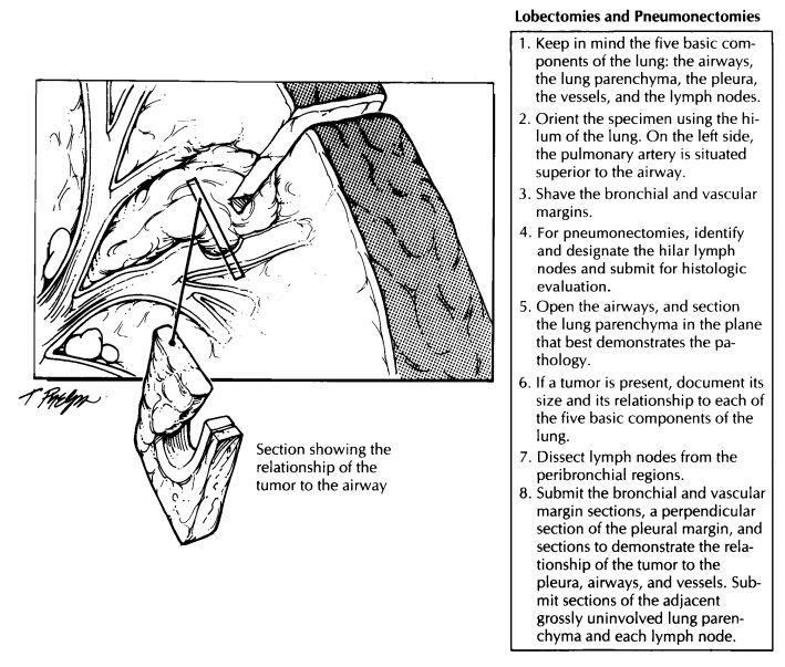
Limited Pulmonary Resections
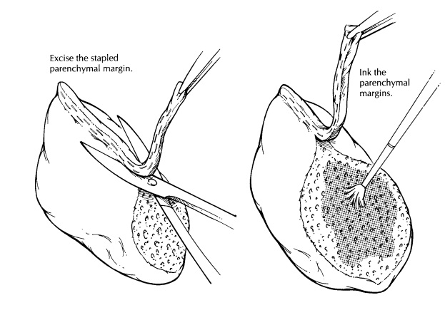
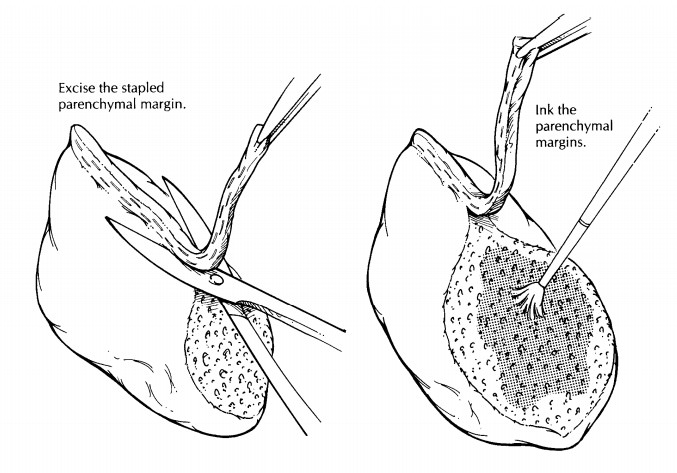
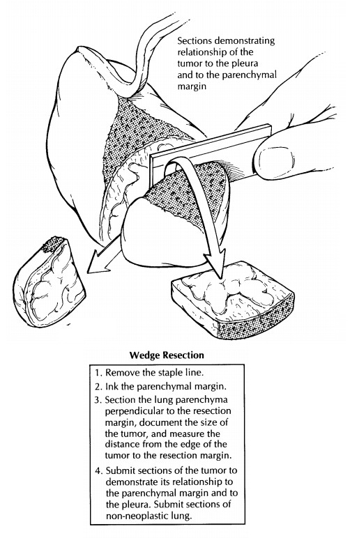
Limited pulmonary resections include open lung biopsies and wedge resections for both neoplastic and non-neoplastic diseases. These specimens are generally taken from the periphery of the lung. As illustrated, they usually are wedge-shaped pieces of lung tissue invested by visceral pleura. Because of their small size and peripheral lo-cation, lymph nodes and major bronchi are usu-ally not present. Surgical staple lines may be present, and these represent the parenchymal resection margins. Document the dimensions of the specimen and the appearance of the pleural surface. If clinically indicated, fresh tissue can be harvested for microbial cultures and for im-munofluorescence. If immediate dissection is not required, however, fix the specimen before pro-ceeding with the dissection. Fixation in distention can be accomplished by gently infusing forma-lin directly into the lung parenchyma at several sites through a small-gauge needle. Take care not to overdistend the specimen. Submerge the dis-tended specimen in formalin until it is well fixed. Following fixation, trim the staple lines from the specimen. Be careful not to remove too much lung tissue with these staples, because the ex-posed lung parenchyma immediately adjacent to the staples represents a surgical margin. Dry and ink the exposed parenchymal margin, and then serially section the specimen in a plane per-pendicular to that of the parenchymal resection margin.
After
sectioning the specimen, evaluate the lung for focal and diffuse processes, and
describe these findings. For neoplasms, note the size of the tumor, the
appearance of its cut surface, and the relationship of the tumor to the pleura
and to the surrounding lung parenchyma. Note also the distance from the edge of
the tumor to the resection margin. Submit one to four sections of the lesion
(depending on its size), selecting sec-tions that demonstrate the tumor’s relationship
to the pleura, to adjacent lung tissue, and to the parenchymal resection
margin. Sample the parenchymal margin, using perpendicular sec-tions when the
tumor closely approaches the margin. In addition, submit two sections of
non-neoplastic lung. Intrapulmonary lymph nodes and bronchi will generally not
be present in these peripheral lung biopsies, but if they are present, they
should be sampled.
For
non-neoplastic lung diseases, submit the vast majority of the specimen for
histologic evaluation. In selected instances, a representative section should
be fresh-frozen, especially if im-munofluorescence studies are needed to
establish a diagnosis. Note the size of the airspaces, the patency of any
airways that are present, and the crepitance of the parenchyma. Only rarely
should intraoperative frozen section consultations be performed for
non-neoplastic lung disease.
Related Topics