Chapter: Human Neuroanatomy(Fundamental and Clinical): Gross Anatomy of the Cerebral Hemispheres
Inferior Surface of Cerebrum - Gross Anatomy of The Cerebral Hemispheres
Gross Anatomy of The Cerebral Hemispheres
Inferior Surface of Cerebrum
When the cerebrum is separated from the hindbrain by cutting across the midbrain, and is viewed from below, the appearances seen are shown in Fig. 8.7. Posterior to the midbrain we see the under surface of the splenium of the corpus callosum. Anterior to the midbrain there is a depressed area
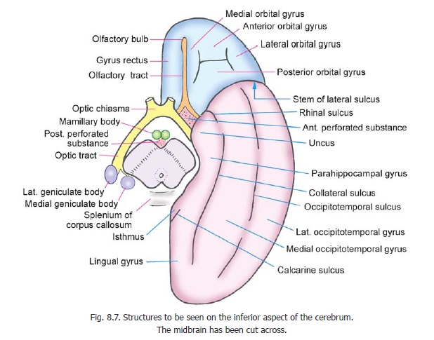
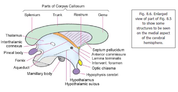
called the interpeduncular fossa. The fossa is bounded in front by the optic chiasma and on the sides by the right and left optic tracts. The optic tracts wind round the sides of the midbrain to terminate on its posterolateral aspect. In this region two swellings, themedial and lateral geniculatebodies, can be seen. Certain structures are seen within the interpeduncular fossa. These are closelyrelated to the floor of the third ventricle (see also Fig. 8.6). Anterior and medial to the crura of the midbrain there are two rounded swellings called the mamillary bodies. Anterior to these bodies there is a median elevation called the tuber cinereum, to which the infundibulum of the hypophysis cerebri is attached. The triangular interval between the mamillary bodies and the midbrain is pierced by numerous small blood vessels and is called the posterior perforated substance. A similar area lying on each side of the optic chiasma is called the anterior perforated substance. The anterior perforated substance is bounded anterolaterally by the lateral olfactory stria and posterolaterally by the uncus. The anterior perforated substance is connected to the insula by a band of grey matter called the limen insulae which lies in the depth of the stem of the lateral sulcus.
In addition to these structures we see the sulci and gyri on the orbital and tentorial parts of the inferior surface of the each cerebral hemisphere. These parts are separated from each other by the stem of the lateral sulcus.
Sulci and gyri on orbital surface
Close to the medial border of the orbital surface there is an anteroposterior sulcus: it is called the olfactory sulcus because the olfactory bulb and tract lie superficial to it. The area medial to this sulcus is called the gyrus rectus. The rest of the orbital surface is divided by an H-shaped orbitalsulcus into anterior, posterior, medial and lateral orbital gyri.
Sulci and gyri on tentorial surface
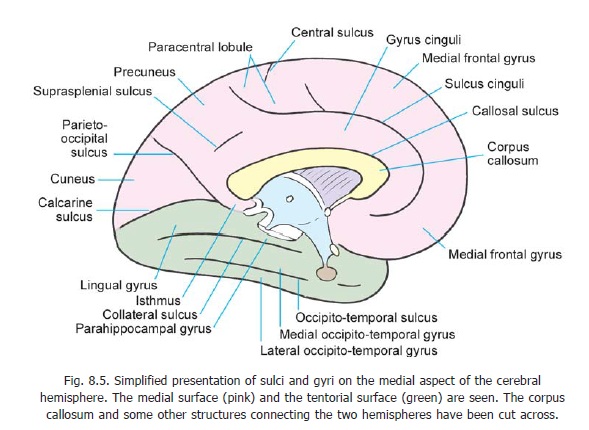
The tentorial surface is marked by two major sulci that run in an anteroposterior direction. These are the collateral sulcusmedially, and the occipito-temporal sulcus laterally. The posterior part of the collateral sulcus runs parallel to the calcarine sulcus: the area between them is the lingualgyrus. Anteriorly, the lingual gyrus becomes continuous with the parahippocampal gyrus which isrelated medially to the midbrain and to the interpeduncular fossa. The anterior end of the parahippocampal gyrus is cut off from the curved temporal pole of the hemisphere by a curved rhinalsulcus. This part of the parahippocampal gyrus forms a hook-like structure called the uncus, detailsof which are considered later. Posteriorly, the parahippocampal gyrus becomes continuous with the gyrus cinguli through the isthmus (Fig. 8.5). The area between the collateral sulcus and the rhinal sulcus medially, and the occipitotemporal sulcus laterally, is the medial occipitotemporal gyrus. The area lateral to the occipitotemporal sulcus is called the lateral occipitotemporal gyrus. This gyrus is continuous (around the inferolateral margin of the cerebral hemisphere) with the inferior temporal gyrus.
AN INTRODUCTION TO SOME STRUCTURES WITHIN THE CEREBRAL HEMISPHERES
The surface of the cerebral hemisphere is covered by a thin layer of grey matter called the cerebralcortex. The cortex follows the irregular contour of the sulci and gyri of the hemisphere and extendsinto the depths of the sulci. As a result of this folding of the cerebral surface, the cerebral cortex acquires a much larger surface area than the size of the hemispheres would otherwise allow.
The greater part of the cerebral hemisphere deep to the cortex is occupied by white matter within which are embedded certain important masses of grey matter. Immediately lateral to the third ventricle
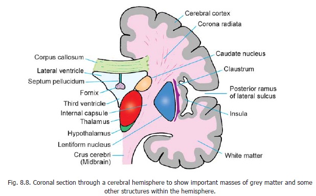
there are the thalamus and hypothalamus (and certain smaller masses) derived from the diencephalon. More laterally there is thecorpus striatum which is derived from the telecephalon. It consists of two masses of grey matter, the caudate nucleus and thelentiform nucleus. A little lateral to the lentiform nucleus we see the cerebral cortex in the region of the insula. Between the lentiform nucleus and the insula there is a thin layer of grey matter called the claustrum. The caudate nucleus, the lentiform nucleus, the claustrum and some other masses of grey matter (all of telencephalic origin) are referred to as basal nuclei or as basal ganglia.
The white matter that occupies the interval between the thalamus and caudate nucleus medially, and the lentiform nucleus laterally, is called the internal capsule. It is a region of considerable importance as major ascending and descending tracts pass through it. The white matter that radiates from the upper end of the internal capsule to the cortex is called the corona radiata.
The two cerebral hemispheres are interconnected by fibres passing from one to the other. These fibres constitute thecommissures of the cerebrum. The largest of these is the corpus callosum which is seen just above the lateral ventricles in Fig. 8.8.
Related Topics