Chapter: Human Neuroanatomy(Fundamental and Clinical): Gross Anatomy of the Cerebral Hemispheres
Exterior of the Cerebral Hemispheres - Gross Anatomy of The Cerebral Hemispheres
Gross Anatomy of The Cerebral Hemispheres
Exterior of the Cerebral Hemispheres
Poles, Surfaces, and Borders
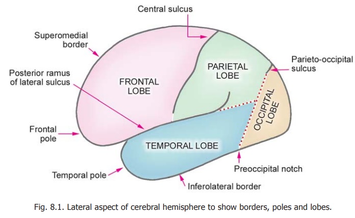
The cerebrum consists of two cerebral hemispheres that are partially connected with each other. When viewed from the lateral aspect each cerebral hemisphere has the appearance shown in Fig. 8.1. Three somewhat pointed ends or poles can be recognised. These are the frontal pole anteriorly, the occipital pole posteriorly, and the temporal pole that lies between the frontal and occipital poles,and points forwards and somewhat downwards.
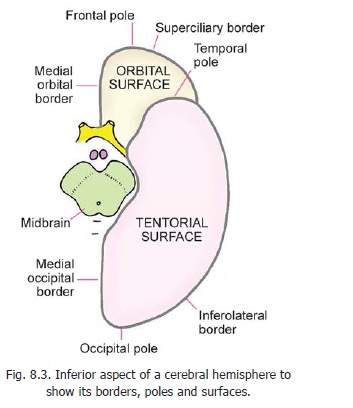
A coronal section through the cerebral hemispheres (Fig. 8.2) shows that each hemisphere has three borders: superomedial, inferolateral and inferomedial. These borders divide the surface of the hemisphere into three large surfaces: superolateral, medialand inferior. The inferior surface is further subdivided into an anterior orbital part and a posterior tentorial part. (Fig. 8.3).
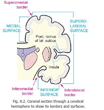
Corresponding to these subdivisions, the inferomedial border is divided into an anterior part called the medial orbital border and a posterior part called the medial occipital border. The orbital part of the inferolateral border is called the superciliary border (as it lies just above the level of the eyebrows).
The surfaces of the cerebral hemisphere are not smooth. They show a series of grooves or sulci which are separated by intervening areas that are called gyri.
Lobes
For convenience of description each cerebral hemisphere is divided into four major subdivisions or lobes. To consider the boundaries of these lobes reference has to be made to some sulci and otherfeatures to be seen on each hemisphere (Fig. 8.1).
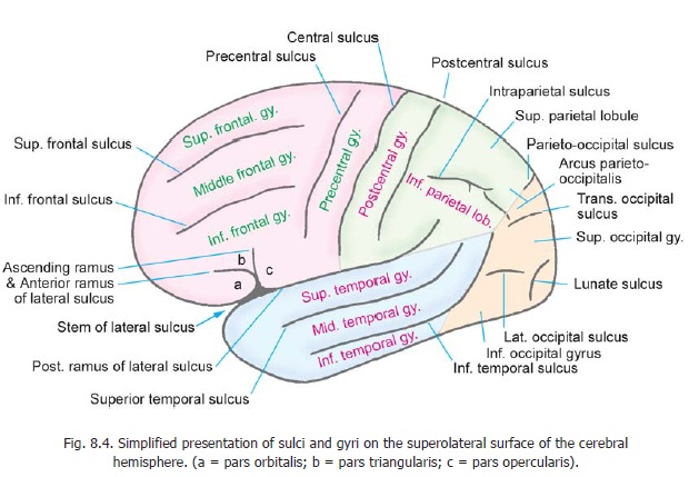
a. On the superolateral surface of the hemisphere there are two prominent sulci. One of these is the posterior ramus of the lateralsulcus which begins near the temporal pole andruns backwards and slightly upwards. Its posteriormost part curves sharply upwards. The second sulcus that is used to delimit the lobes is the central sulcus. It begins on the superomedial margin a little behind the midpoint between the frontal and occipital poles, and runs downwards and forwards to end a little above the posterior ramus of the lateral sulcus.
b. On the medial surface of the hemisphere, near the occipital pole, there is a sulcus called the parietooccipital sulcus (Fig. 8.5). The upperend of this sulcus reaches the superomedial border and a small part of it can be seen on the superolateral surface (Fig. 8.1).
c. A little anterior to the occipital pole the inferolateral border shows a slight indentation called the preoccipital notch (or preoccipital incisure).
To complete the subdivision of the hemisphere into lobes we now have to draw two imaginary lines. The first imaginary line connects the upper end of the parieto-occipital sulcus to the preoccipital notch. The second imaginary line is a backward continuation of the posterior ramus of the lateral sulcus (excluding the posterior upturned part) to meet the first line. We are now in a position to define the limits of the various lobes as follows.
1. The frontal lobe lies anterior to the central sulcus, and above the posterior ramus of the lateral sulcus.
2. The parietal lobe lies behind the central sulcus. It is bounded below by the posterior ramus of the lateral sulcus and by the second imaginary line; and behind by the upper part of the first imaginary line.
3. The occipital lobe is the area lying behind the first imaginary line.
4. The temporal lobe lies below the posterior ramus of the lateral sulcus and the second imaginary line. It is separated from the occipital lobe by the lower part of the first imaginary line.
Before going on to consider further subdivisions of each of the lobes named above, attention has to be directed to details of some structures already mentioned.
a. The upper end of the central sulcus winds round the superomedial border to reach the medial surface. Here its end is surrounded by a gyrus called the paracentral lobule (Fig. 8.5). The lower end of the central sulcus is always separated by a small interval from the posterior ramus of the lateral sulcus (Fig. 8.1).
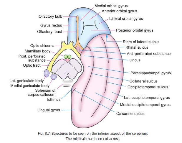
b. The lateral sulcus begins on the inferior aspect of the cerebral hemisphere where it lies between the orbital surface and the anterior part of the temporal lobe (Fig. 8.7). It runs laterally to reach the superolateral surface. On reaching this surface it divides into three rami (branches). These rami are anterior (or anterior horizontal), ascending (or anterior ascending) and posterior (Fig.8.4). The anterior and ascending rami are short and run into the frontal lobe in the directions indicated by their names. The posterior ramus has already been considered. Unlike most other sulci, the lateral sulcus is very deep. Its walls cover a fairly large area of the surface of the hemisphere called the insula (Fig. 8.2).
Further Subdivisions of the Superolateral Surface
The subdivisions of the superolateral surface are described below and are shown in Fig. 8.4.
Frontal lobe
The frontal lobe is further subdivided as follows. The precentral sulcus runs downwards and forwards parallel to and a little anterior to the central sulcus. The area between it and the central sulcus is the precentral gyrus. In the region anterior to the precentral gyrus there are two sulci that run in an anteroposterior direction. These are the superior and inferior frontal sulci. They dividethis region into superior, middle and inferior frontal gyri. The anterior and ascending rami of the lateral sulcus extend into the inferior frontalgyrus dividing it into three parts. The part below the anterior ramus is the pars orbitalis; that between the anterior and ascending rami is the pars triangularis; and the part posterior to the ascending ramus is the pars opercularis.
Temporal lobe
The temporal lobe has two sulci that run parallel to the posterior ramus of the lateral sulcus. They are termed the superior and inferior temporal sulci. They divide the superolateral surface of this lobe into superior, middle and inferior temporal gyri.
Parietal lobe
The parietal lobe shows the following subdivisions. The postcentral sulcus runs downwards and forwards parallel to and a little behind the central sulcus. The area between these two sulci is the postcentral gyrus. The rest of the parietal lobe is divided into asuperior parietal lobule and an inferior parietal lobule by the intraparietal sulcus. The upturned posterior end of the posteriorramus of the lateral sulcus extends into the inferior parietal lobule. The posterior ends of the superior and inferior temporal sulci also turn upwards to enter this lobule. The upturned ends of these three sulci divide the inferior parietal lobule into three parts. The part that arches over the upturned posterior end of the posterior ramus of the lateral sulcus is called the supramarginal gyrus. The part that arches over the superior temporal sulcus is called the angular gyrus. The part that arches over the posterior end of the inferior temporal sulcus is called the arcus temporooccipitalis.
Occipital lobe
The occipital lobe shows three rather short sulci. One of these, the lateral occipital sulcus lies horizontally and divides the lobe into superior and inferior occipital gyri. The lunate sulcus runs downwards and slightly forwards just in front of the occipital pole. The vertical strip just in front of it is the gyrus descendens. The transverse occipital sulcus is located in the uppermost part of the occipital lobe. The upper end of the parieto-occipital sulcus (which just reaches the superolateral surface from the medial surface) is surrounded by the arcus parieto-occipitalis. As its name suggests, it belongs partly to the parietal lobe and partly to the occipital lobe.
Insula
In the depth of the stem and posterior ramus of the lateral sulcus there is a part of the cerebral hemisphere called the insula (insula = hidden). It is surrounded by a circular sulcus. During development of the cerebral hemisphere this area grows less than surrounding areas which, therefore, come to overlap it and occlude it from surface view. These surrounding areas are called opercula(= lids). The frontal operculum lies between the anterior and ascending rami of the lateral sulcus. The frontoparietal operculum lies above the posterior ramus of the lateral sulcus. The temporal operculum lies below this sulcus. The temporal operculum has a superior surface hidden in the depthof the lateral sulcus (Figs. 8.2, 8.15B). On this surface are located two gyri called the anterior andposterior transverse temporal gyri. The surface of the insula itself is divided into a number ofgyri.
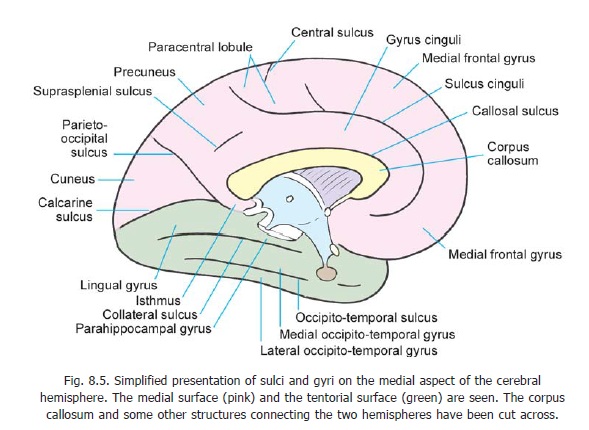
Medial Surface of Cerebral Hemisphere
When the two cerebral hemispheres are separated from each other by a cut in the midline the appearances seen are shown in Figs. 8.5 and 8.6.
The structures seen are as follows.
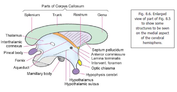
The corpus callosum is a prominent arched structure consisting of commissural fibres passing from one hemisphere to the other (Fig. 8.6). It consists of a central part called the trunk, a posterior end or splenium, and an anterior end or genu. A little below the corpus callosum we see the third
ventricle of the brain. A number of structures can be identified in relation to this ventricle. The interventricular foramen through which the third ventricle communicates with the lateral ventriclecan be seen in the upper and anterior part. Posteroinferiorly, the ventricle is continuous with the cerebral aqueduct. The lateral wall of the ventricle is formed in greater part by a large mass ofgrey matter called the thalamus. The right and left thalami are usually interconnected (across the midline) by a strip of grey matter called the interthalamic connexus. The anteroinferior part of the lateral wall of the third ventricle is formed by a collection of grey matter that constitutes the hypothalamus.
Above the thalamus there is a bundle of fibres called the fornix. Posteriorly, the fornix is attached to the under surface of the corpus callosum, but anteriorly it disappears from view just in front of the interventricular foramen. Extending between the fornix and the corpus callosum there is a thin lamina called the septum pellucidum (or septum lucidum), which separates the right and left lateral ventricles from each other. Removal of the septum pellucidum brings the interior of the lateral ventricle into view.
In the anterior wall of the third ventricle there are the anterior commissure and the laminaterminalis. The anterior commissure is attached to the genu of the corpus callosum through a thinlamina of fibres that constitutes the rostrum of the corpus callosum. Below, the anterior commissure is continuous with the lamina terminalis which is a thin lamina of nervous tissue. The lower end of the lamina terminalis is attached to the optic chiasma. Just in front of the lamina terminalis there are the paraterminal gyrus and theparolfactory gyrus (Fig. 16.10). Posteriorly, the third ventricle is related to the pineal body (or pineal gland) and inferiorly to thehypophysis cerebri.
Above the corpus callosum (and also in front of and behind it) we see the sulci and gyri of the medial surface of the hemisphere (Fig. 8.5). The most prominent of the sulci is the cingulate sulcus which follows a curved course parallel to the upper convex margin of the corpus callosum. Anteriorly, it ends below the rostrum of the corpus callosum. Posteriorly, it turns upwards to reach the superomedial border a little behind the upper end of the central sulcus. The area between the cingulate sulcus and the corpus callosum is called the gyrus cinguli. It is separated from the corpus callosum by the callosal sulcus.
The part of the medial surface of the hemisphere between the cingulate sulcus and the superomedial border consists of two parts. The smaller posterior part which is wound around the end of the central sulcus is called the paracentral lobule. The large anterior part is called the medial frontal gyrus. These two parts are separated by a short sulcus continuous with the cingulate sulcus.
The part of the medial surface behind the paracentral lobule and the gyrus cinguli shows two major sulci that cut off a triangular area called the cuneus. The triangle is bounded anteriorly and above by the parieto-occipital sulcus; inferiorly by the calcarine sulcus; and posteriorly by the superomedial border of the hemisphere. The calcarine sulcus extends forwards beyond its junction with the parieto-occipital sulcus and ends a little below the splenium of the corpus callosum. The small area separating the splenium from the calcarine sulcus is called the isthmus. Between the parieto-occipital sulcus and the paracentral lobule there is a quadrilateral area called the precuneus. Anteroinferiorly the precuneus is separated from the posterior part of the gyrus cinguli by the suprasplenial (orsubparietal) sulcus.
The precuneus and the posterior part of the paracentral lobule form the medial surface of the parietal lobe.
Advanced:
Although the parieto-occipital and calcarine sulci appear to be continuous with each other on surface view, they are separated by the cuneate gyrus (or cuneolingual gyrus) which lies in the depth of the area where the two sulci meet. The parts of the calcarine sulcus anterior and posterior to the junction with the parieto-occipital sulcus are separated by a deeply situated anterior cuneolingualgyrus.
Related Topics