Chapter: Ophthalmology: Eye Lens
Eye Lens: Basic Knowledge
Lens
Basic Knowledge
Function of the lens:
The lens is one of the essential refractive media of theeye and
focuses incident rays of light on the retina. It adds a variable element to the
eye’s total refractive power (10 – 20 diopters, depending on individual
accommodation) to the fixed refractive power of the cornea (approximately 43
diopters).
Shape:
The fully developed lens is abiconvex,transparent
structure. Thecurvature of the posterior surface, which has a radius of 6
mm, is greater than that of the anterior surface, which has a radius of 10 mm.
Weight:
The lens is approximately 4 mm thick, and its weight increases
withage to five times its weight at birth. An adult lens weighs about 220 mg.
Position and suspension:
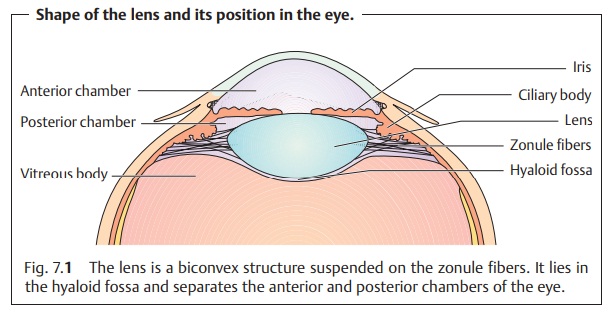
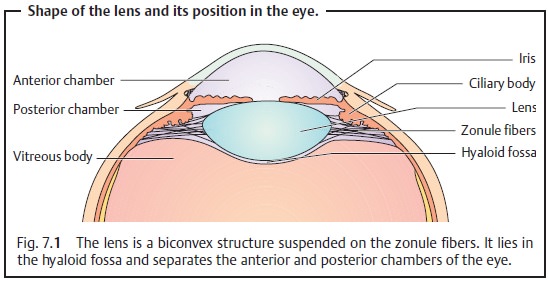
The lens lies in the posterior chamber of the eyebetween the
posterior surface of the iris and the vitreous body in a saucer-shaped depression of the vitreous body known as the hyaloid
fossa. Togetherwith the iris it forms an optical diaphragm that separates the
anterior and posterior chambers of the eye. Radially arranged zonule fibers that insert into the lens
around its equator connect the lens to the ciliary body. These fibers hold the
lens in position (Fig. 7.1) and
transfer the tensile force of the ciliary muscle (see Accommodation).
Embryology and growth:
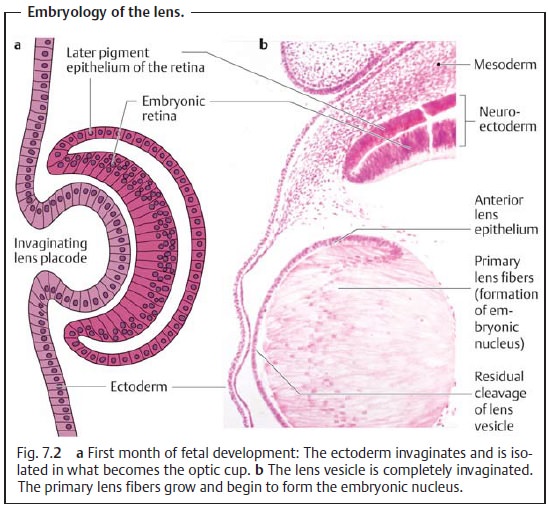
The lens is apurely epithelial structurewithoutany nerves or blood vessels. It moves into its intraocular position in the first month of fetal development as surface ectoderm invaginates into the primi-tive optic vesicle, which consists of neuroectoderm. A purely ectodermal struc-ture, the lens differentiates during gestation into central geometric lensfibers, an anterior layer of epithelial cells, and an acellular hyaline capsule (Figs. 7.2a and b). The normal direction of growth of epithelial structures is centrifugal; fully developed epithelial cells migrate to the surface and are peeled off. However, the lens grows in the opposite direction. The youngest cells are always on the surface and the oldest cells in the center of the lens. The growth of primary lens fibers forms the embryonic nucleus. At the equa-tor, the epithelial cells further differentiate into lens fiber cells (Fig. 7.2).
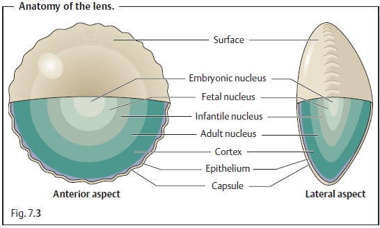
These new secondary fibers displace the
primary fibers toward the center of the lens. Formation of a fetal nucleus that encloses the embryonic nucleus is complete at birth. Fiber
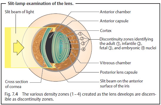
formation at the equator, which continues through-out life, produces the infantile nucleus during the first and second decades of life, and the adult nucleus during the third decade. Completely enclosed by the lens
capsule, the lens never loses any cells so that its tissue is continuously
compressed throughout life (Fig. 7.3a and b). The various density zones created as the lens develops are
readily discernible as discontinuity zones (Fig. 7.4).
Metabolism and aging of the lens:
The lens is nourished bydiffusion
fromthe aqueous humor. In this respect it resembles a tissue culture, with
theaqueous humor as its substrate and the eyeball as the container that
provides a constant temperature.
The metabolism and detailed biochemical
processes involved in aging are complex and not completely understood. Because
of this, it has not been possible to influence cataract development with medications.
The metabolism and growth of the lens cells
are self-regulating. Metabolic activity is essential for the preservation of
the integrity, transparency, and optical function of the lens. The epithelium
of the lens helps to maintain the ion equilibrium and permit transportation of nutrients, minerals, and waterinto the lens. This type of transportation, referred to as
a “pump-leak sys-tem,” permits
active transfer of sodium, potassium, calcium, and amino acids
Maintaining this
equilibrium (homeostasis) is essen-tial for the transparency of the lens and is
closely related to the water balance. The water content of the lens is normally stable and in equilibrium with
the surrounding aqueous humor. The water content of the lens decreases with
age, whereas the content of insoluble lens proteins (albuminoid) increases. The
lens becomes harder, less elastic (see Loss of accommodation), and less
transparent. A decrease in the transparency of the lens with age is as unavoidable as wrinkles in the skin or
gray hair. Manifestly reduced trans-parency is present in 95% of all persons
over the age of 65, although individual exceptions are not uncommon. The
central portion or nucleus of the lens becomes sclerosed and slightly yellowish
with age.
Related Topics