Chapter: Ophthalmology: The Ophthalmic Examination
Examination of the Cornea
Examination of the Cornea
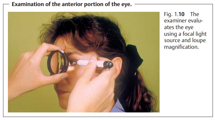
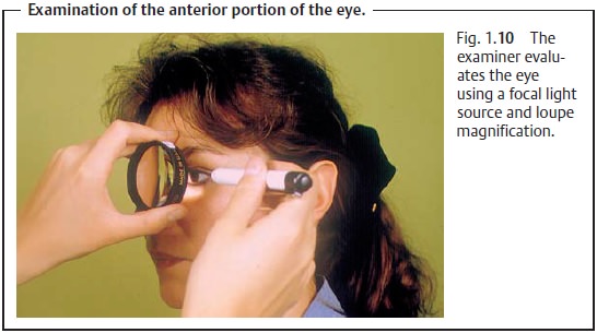
The cornea is examined with a point light source and a loupe (Fig. 1.10). The cornea is smooth,
clear, and reflective. The reflection is distorted in the pres-ence of corneal
disorders. Epithelial defects, which are also very painful, will take on an
intense green color after application of fluorescein dye; corneal infiltrates
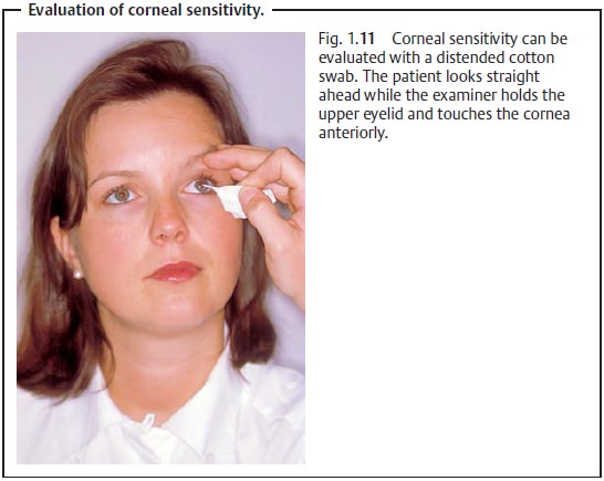
and scars are grayish white. Evaluating corneal sensitivity is also important.
Sensitivity is evaluated bilaterally to detect possible differences in the
reaction of both eyes. The patient looks straight ahead during the
exami-nation. The examiner holds the upper eyelid to prevent reflexive closing
and touches the cornea anteriorly (Fig. 1.11). Decreased sensitivity can provide
information about trigeminal or facial neuropathy, or may be a sign of a viral
infection of the cornea.
Related Topics