Chapter: Microbiology and Immunology: Virology, Virus: Parvoviruses
Coxsackieviruses
Coxsackieviruses
Coxsackieviruses are so named, because the viruses were first isolated in Coxsackie village in New York, United States, by Dandruff and Sickel in the year 1985. These viruses based on the pathological changes produced in suckling mice are classified into two groups: coxsackieviruses A and coxsackieviruses B.
The structure and morphology of the coxsackieviruses and the RNA genome are similar to those of poliovirus. But unlike poliovirus, they can infect mammals other than primates. Replication cycle is similar to that of poliovirus.
Coxsackie A viruses consist of 24 serotypes and are associated with diseases involving vesicular lesions, such as herpangina. Coxsackie B viruses consist of six serotypes and are most frequently associated with myocarditis and pleurodynia.
Coxsackie A viruses show tropism for skin and mucous membranes, whereas coxsackie B viruses show predilection for visceral organs, such as liver, heart, pancreas, and pleura. Both the viruses infect anterior horn cells of the motor neurons and meninges and cause paralysis. They replicate first in the oropharynx and gastrointestinal tract from where they spread by the blood circulation. Host immunity is IgG-antibody mediated and is type specific.
Coxsackievirus infection occurs worldwide. Humans are the only natural hosts. The viruses are transmitted primarily by fecal–oral route.
Diseases Caused by Coxsackie A
Diseases specifically caused by coxsackie A viruses include the following:
Herpangina: This condition is caused by coxsackie A virusserotypes 2, 4, 5, 6, 8, and 10. The infection is most commonly seen in children between 1 and 7 years. The symptoms include sudden onset of fever, sore throat, and difficulty in swallow-ing. The classical finding is a painful vesicular eruption of the oral mucosa around the soft palate and uvula. Less commonly, these lesions may be found on the hard palate. The condition is self-limiting. The viruses are isolated from the lesions or from the feces.
Hand-foot-and-mouth disease: This is a vesicular exan-thema usually caused by coxsackievirus serotypes 5 and 16. This is mainly a disease of children, seen most commonly in patients younger than 10 years. After an incubation period of 3–6 days, the infection begins with prodromal symptoms, such as fever, cough, sore throat, malaise, and anorexia. After 12–36 hours, vesicular eruptions appear on the hands, feet, and oral cavity. This is a self-limiting condition and the illness subsides in a few days.
Diseases Caused by Coxsackie B
Diseases specifically caused by coxsackie B viruses include the following:
Pleurodynia: This condition, also known as epidemic myalgiaor Bornholm disease, is caused by coxsackie B virus serotypes 3 and 5. This condition is an acute illness, which manifests with a sudden onset of fever accompanied by muscular pain and pain in the chest and abdomen. The pain is spasmodic in nature. The condition usually lasts for 4 days but may relapse again after the condition has been asymptomatic for several days.
Myocarditis: This is a serious condition caused by coxsackieB virus, mostly in newborn infants. Shortness of breath, dull or sharp chest pain, and fever are the common manifestations of the condition. This is a life-threatening condition mostly in newborns. In adults, the condition presents with myocardial infection with fever. The condition is associated with a high morbidity.
Diseases Caused by Coxsackie A and B
Diseases caused by both coxsackie A and B viruses include aseptic meningitis.
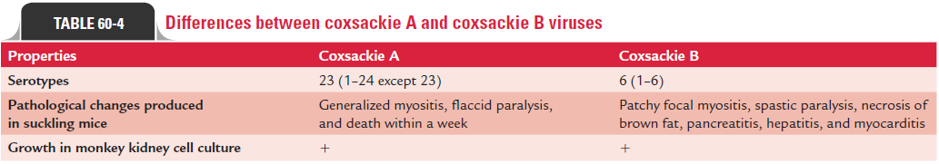
Aseptic meningitis: The condition is caused by coxsackieA serotypes 2, 4, 7, and 9 and by coxsackie B viruses. The labora-tory diagnosis of coxsackievirus infection is made by isolation of the virus from the feces or from lesions by intracerebral or intra-peritoneal inoculation into a 1-day-old mouse. Identification of the virus is carried out by histopathology study of the infected mouse after sacrifice. Some of the coxsackieviruses can also be isolated by culture in human diploid embryonic lung fibro-blasts, BGM, and HeLa cell lines. Cytopathic effects resemble those of polioviruses but develop more slowly, beginning as foci of rounded refractile cells, which die and fall off the glass. Differences between coxsackieviruses A and B are summarized in Table 60-4.
Related Topics