Chapter: Medicine and surgery: Dermatology and soft tissues
Clinical - Dermatology and soft tissues
Clinical
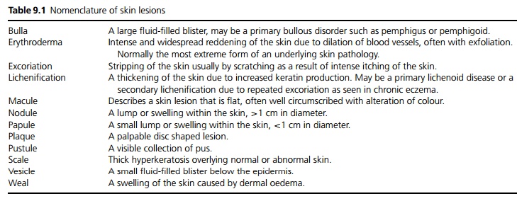
Nomenclature and description
The cornerstone of dermatological diagnosis is accurate observation and description of lesions and rashes. Some terms used to describe specific lesions are given in Table 9.1. Many rashes have classical distributions, which may be symmetrical or asymmetrical. Specific rashes and distributions are considered under individual headings.
Dermatological procedures
· Shave or tangential excision: This procedure slices a surface growth off using a blade, often to remove a small growth and confirm its nature at the same time.
· Punch biopsy: Under local anaesthesia a full thickness cylinder of skin (1–4 mm diameter) is cut out using a biopsy punch. The resultant hole is sutured and leaves minimal scarring.
· Electrodesiccation and curettage: Under local anaesthesia lesions including precancers and benign growths are scraped off with a special tool and the area is cauterised to stop bleeding. Repeated treatment may be required. The area heals often leaving a small hypopigmented mark.
· Cryotherapy: Liquid nitrogen is used to freeze the cutaneous lesion. Light freezing causes a peeling, moderate freezing a blistering and hard freezing a scabbing.
· Mohs’ surgery: This is a technique used in the resection of basal cell and squamous cell carcinomas.
Under local anaesthesia palpable tumour is excised with a curette or scalpel. A thin section a few millimetres around and underneath the resulting defect is taken, divided into pieces, and cut as a fresh frozen specimen. If tumour is seen at a particular margin resection is continued at the appropriate margin, and further sections examined until no further tumour is seen. This technique allows the maximal conservation of normal surrounding tissue. If the resultant defect is large, formal reconstructive surgery may be necessary.
Skin grafts
Skin grafts are sections of skin that are completely detached and transferred to cover large areas of skin defect. The recipient site requires a good blood supply, as the graft has no supply of its own.
· Split skin grafts are used in acute trauma, granulating areas and burns. A guarded freehand knife or an electric dermatome is used to remove epidermis and a variable amount of dermis from the donor site, which heals by reepithelialisation. If a very large defect needs covering, the graft can be meshed. Split skin grafts take up a blood supply more easily than full thickness grafts, but tend to shrink and have abnormal pigmentation and contour.
· Full thickness grafts, involving epidermis and entire dermis, are used mainly in reconstructive surgery. They leave a donor site, which requires closure by sutures, limiting the size of the graft. They require a very good vascular bed at the recipient site to survive.
Skin flaps
Skin flaps differ from skin grafts in that they are taken with their own blood supply. The coverage can thus be thicker and stronger than grafts, and can be applied to avascular areas such as exposed bone, tendons and joints. Flaps may be transferred whilst maintaining their original vascular attachments (pedicle flaps), or may be reanastamosed to local blood supply (free flaps).
Related Topics