Chapter: Essentials of Anatomy and Physiology: Skeletal System: Bones and Joints
Appendicular Skeleton
APPENDICULAR SKELETON
A.Identify the bones that make up the pectoral girdle, andrelate their structure and arrangement to the function of the girdle.
B. Name and describe the major bones of the upper limb.
C.Name and describe the bones of the pelvic girdle andexplain why the pelvic girdle is more stable than thepectoral girdle.
D.Name the bones that make up the coxal bone. \istinguishbetween the male and female pelvis.
E.Identify and describe the bones of the lower limb
The appendicular (ap′\en-dik′\ū-lăr; appendage) skeleton consists of the bones of the upper and lower limbs, as well as the girdles, which attach the limbs to the axial skeleton.
Pectoral Girdle
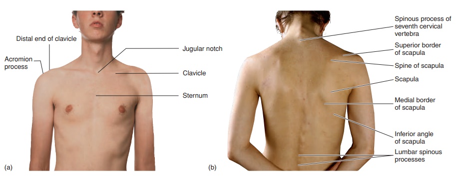
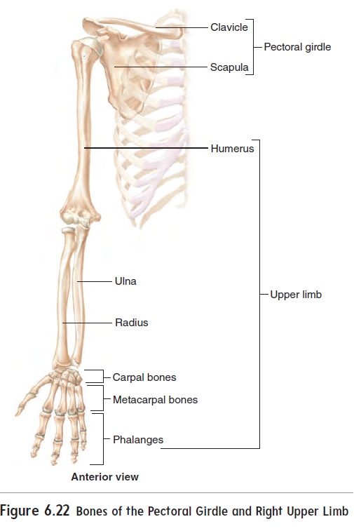
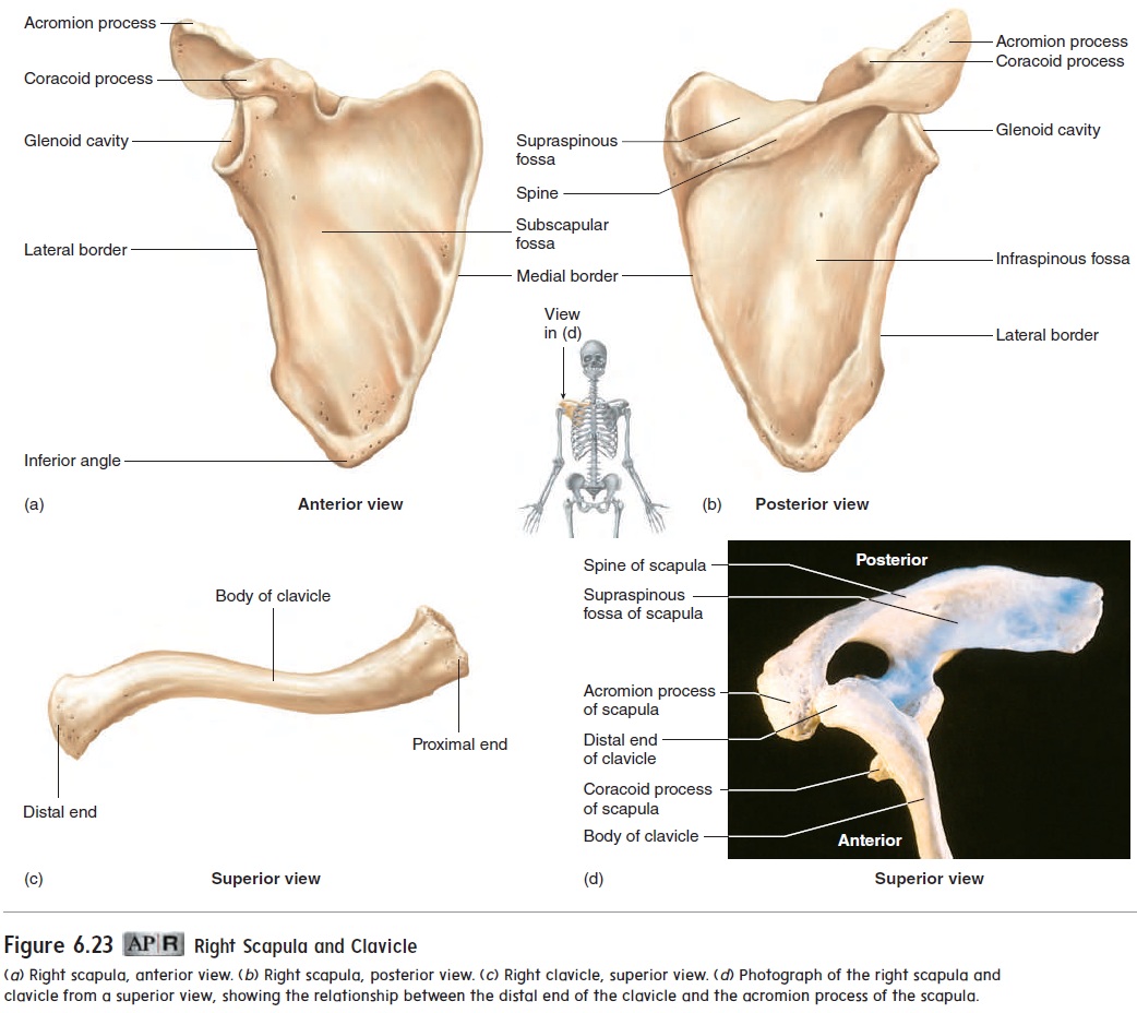
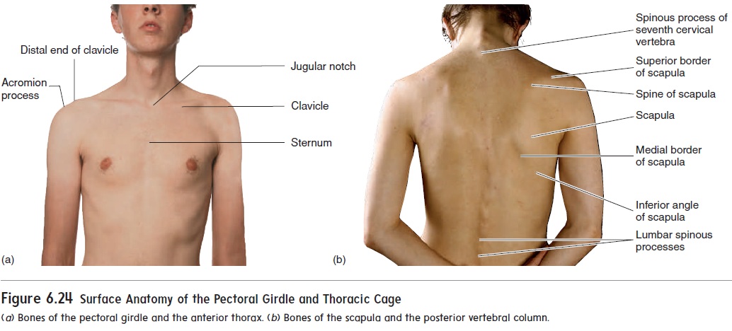
The pectoral (pek′\tō-răl) girdle, or shoulder girdle (figure 6.22), consists of four bones, two scapulae and two clavicles, which attach the upper limb to the body. The scapula (skap′\ū-lă), or shoulder blade, is a flat, triangular bone with three large fossaewhere muscles extending to the arm are attached (figures 6.23 and 6.24; see figure 6.10). A fourth fossa, the glenoid (glen′\oyd) cavity, is where the head of the humerus connects to the scapula.A ridge, called the spine, runs across the posterior surface of the scapula. A projection, called the acromion (ă-krō′\mē-on; akron, tip + omos, shoulder) process, extends from the scapular spine to form the point of the shoulder. The clavicle (klav′\i-kl), or col-larbone, articulates with the scapula at the acromion process. Theproximal end of the clavicle is attached to the sternum, providing the only bony attachment of the scapula to the remainder of the skeleton. The clavicle is the first bone to begin ossification in the fetus. This relatively brittle bone may be fractured in the newborn during delivery. The bone remains slender in children and may be broken as a child attempts to take the impact of a fall on an outstretched hand. The clavicle is thicker in adults and is less vulnerable to fracture. Even though it is the first bone to begin ossification, it is the last to complete ossification. The coracoid (kōr′\ă-koyd) process curves below the clavicle and provides for the attachment of arm and chest muscles.
Upper Limb
The upper limb consists of the bones of the arm, forearm, wrist, and hand (see figure 6.22).
Arm
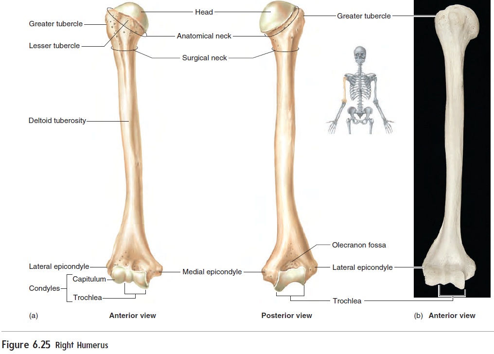
The arm is the region between the shoulder and the elbow; it contains the humerus (hū′\mer-ŭs; shoulder) (figure 6.25). The proximal end of the humerus has a smooth, rounded head, which attaches the humerus to the scapula at the glenoid cavity. Around the edge of the humeral head is the anatomical neck. When the joint needs to be surgically replaced, this neck is not easily accessible. A more accessible site for surgical removal is at the surgical neck, located at the proximal end of the humeral shaft. Lateral to the head are two tubercles, a greater tubercle and a lesser tubercle.Muscles originating on the scapula attach to the greater and lesser tubercles and hold the humerus to the scapula. Approximately one-third of the way down the shaft of the humerus, on the lateral surface, is the deltoid tuberosity, where the deltoid muscle attaches. The size of the deltoid tuberosity can increase as the result of fre-quent and powerful pulls from the deltoid muscle. For example, in bodybuilders, the deltoid muscle and the deltoid tuberosity enlarge substantially. Anthropologists, examining ancient human remains, can use the presence of enlarged deltoid tuberosities as evidence that a person was engaged in lifting heavy objects during life. If the humerus of a person exhibits an unusually large deltoid tuberosity for her age, it may indicate, in some societies, that she was a slave and was required to lift heavy loads. The distal end of the humerus is modified into specialized condyles that connect the humerus to the forearm bones. Epicondyles (ep′\i-kon′\dı̄lz; epi, upon) on the distal end of the humerus, just lateral to the condyles, provide attachment sites for forearm muscles.
Forearm
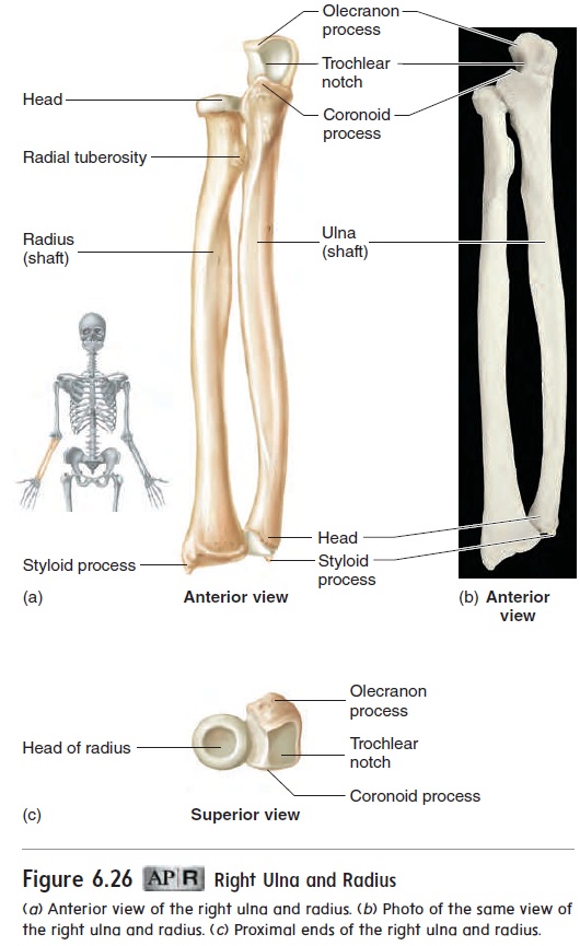
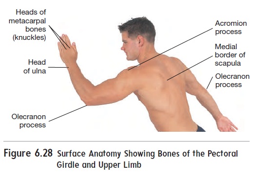
The forearm has two bones: the ulna (ŭl′\nă) on the medial (little finger) side of the forearm and the radius on the lateral (thumb) side (figure 6.26). The proximal end of the ulna forms a trochlearnotch that fits tightly over the end of the humerus, forming most ofthe elbow joint. Just proximal to the trochlear notch is an extension of the ulna, called the olecranon (ō-lek′\ră-non; elbow) process, which can be felt as the point of the elbow (see figure 6.28). Just distal to the trochlear notch is a coronoid (kōr′\ŏ-noyd) process, which helps complete the “grip” of the ulna on the distal end of the humerus. The distal end of the ulna forms a head, which articulates with the bones of the wrist, and a styloid process is located on its medial side. The ulnar head can be seen as a promi-nent lump on the posterior ulnar side of the wrist. The proximal end of the radius has a head by which the radius articulates with both the humerus and the ulna. The radius does not attach as firmly to the humerus as the ulna does. The radial head rotates against the humerus and ulna. Just distal to the radial head is a radial tuberosity, where one of the arm muscles, the biceps bra-chii, attaches. The distal end of the radius articulates with the wrist bones. A styloid process is located on the lateral side of the distal end of the radius. The radial and ulnar styloid processes provide attachment sites for ligaments of the wrist.
Wrist
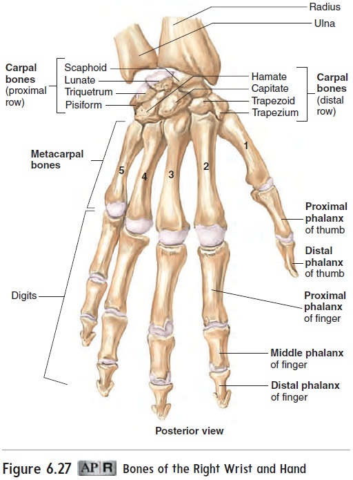
The wrist is a relatively short region between the forearm and the hand; it is composed of eight carpal (kar′\păl; wrist) bones (fig-ure 6.27). These eight bones are the scaphoid (skaf′\oyd), lunate (lū′\nāt), triquetrum (trı̄-kwē′\trŭm), pisiform (pis′\i-fōrm), trape-zium (tra-pē′\zē-ŭm), trapezoid (trap′\ĕ-zoyd), capitate (kap′\i-tāt), and hamate (ha′\māt). The carpal bones are arranged in two rows of four bones each and form a slight curvature that is concave anteriorly and convex posteriorly. A number of mnemonics have been developed to help students remember the carpal bones. The following one allows students to remember them in order from lateral to medial for the proximal row (top) and from medial to lateral (by the thumb) for the distal row: So Long Top Part, Here Comes The Thumb—that is, Scaphoid, Lunate,Triquetrum, Pisiform, Hamate, Capitate, Trapezoid, and Trapezium.
Hand
Five metacarpal (met′\ă-kar′\păl) bones are attached to the carpal bones and form the bony framework of the hand (figure 6.27). The metacarpal bones are aligned with the five digits: the thumb and fingers. They are numbered 1 to 5, from the thumb to the little finger. The ends, or heads, of the five metacarpal bones associated with the thumb and fingers form the knuckles (figure 6.28). Each finger consists of three small bones called phalanges (fă-lan′\jēz; sing. phalanx, fā′\langks), named after the Greek phalanx, a wedge of soldiers holding their spears, tips outward, in front of them. The phalanges of each finger are called proximal, middle, and distal, according to their position in the digit. The thumb has two phalanges, proximal and distal. The digits are also numbered 1 to 5, starting from the thumb.
Pelvic Girdle
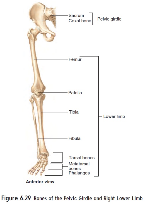
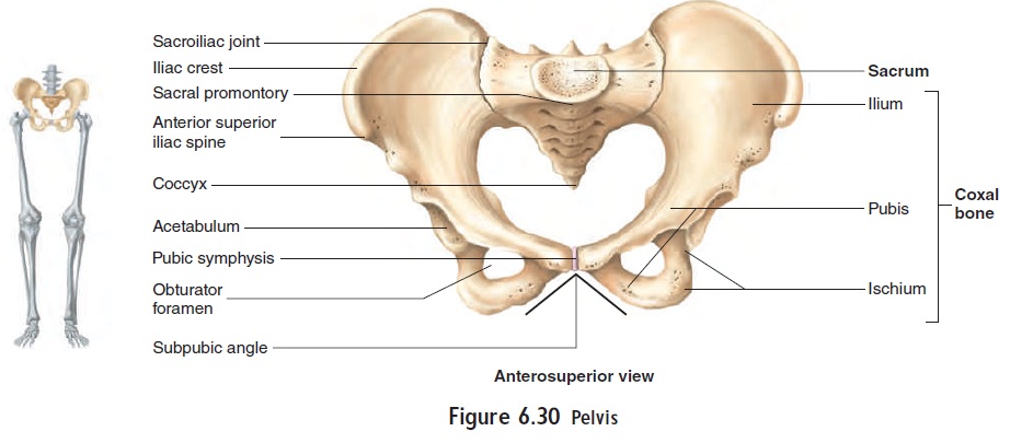
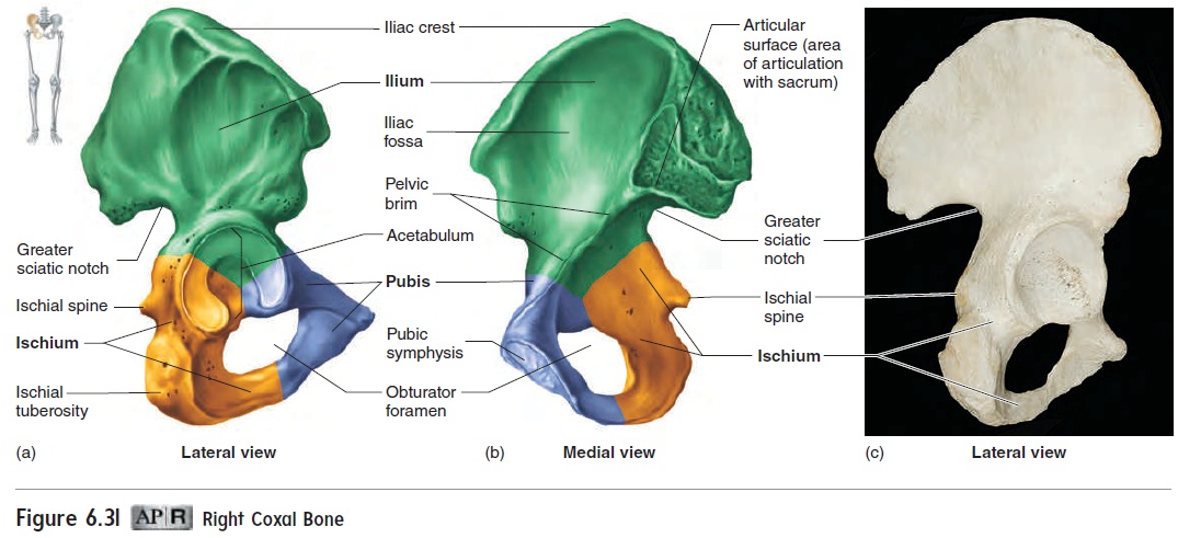
The pelvic girdle is the place where the lower limbs attach to the body (figure 6.29). The right and left coxal (kok′\sul) bones, or hip bones, join each other anteriorly and the sacrum posteriorly to form a ring of bone called the pelvic girdle. The pelvis(pel′\vis; basin) includes the pelvic girdle and the coccyx (figure 6.30). The sacrum and coccyx form part of the pelvis but are also part of the axial skel-eton. Each coxal bone is formed by three bones fused to one another to form a single bone (figure 6.31). The ilium (il′\ē-ŭm) is the most superior, the ischium (is′\kē-ŭm) is inferior and posterior, and the pubis (pū′\bis) is inferior and anterior. An iliac crest can be seen along the superior margin of each ilium, and an anterior superioriliac spine, an important hip landmark, is located at the anterior endof the iliac crest. The coxal bones join each other anteriorly at the pubic(pū′\bik) symphysis and join the sacrum posteriorly at the sacroiliac (sā-krō-il′\ē-ak) joints (see figure 6.30). The acetabulum (as-ĕ-tab′\ū-lŭm; vinegar cup) is the socket of the hip joint. The obturator (ob′\too-rā-tŏr) foramen is the large hole in each coxalbone that is closed off by muscles and other structures.
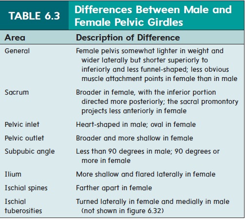
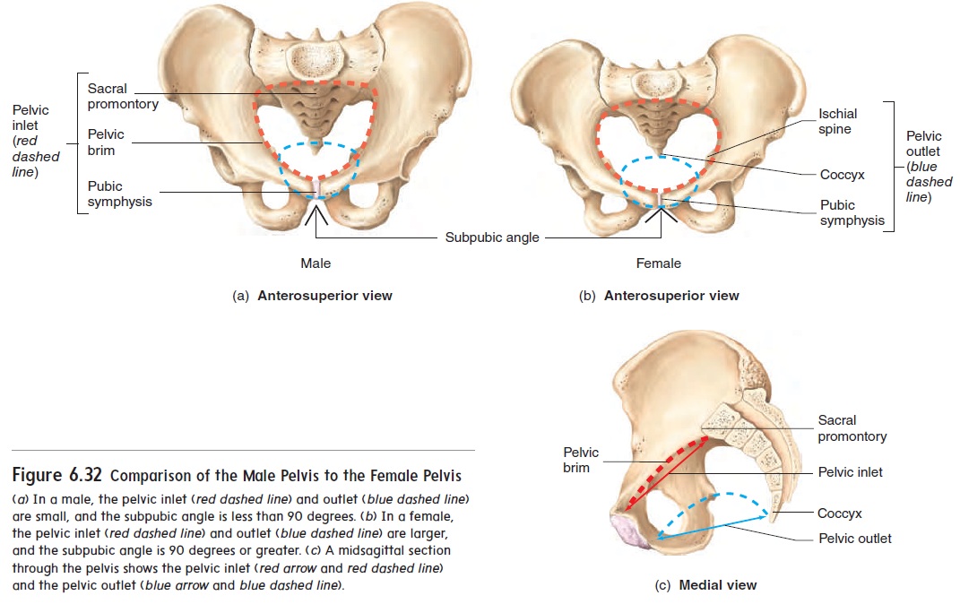
The male pelvis can be distinguished from the female pelvis because it is usually larger and more massive, but the female pelvis tends to be broader (figure 6.32; table 6.3). Both the inlet and the outlet of the female pelvis are larger than those of the male pelvis, and the subpubic angle is greater in the female (figure 6.32a,b). The increased size of these openings helps accommodate the fetus during childbirth. Thepelvic inlet is formed by the pelvic brim and the sacral promontory. The pelvic outlet is bounded by the ischial spines, the pubic symphysis, and the coccyx (figure 6.32c).
Lower Limb
The lower limb consists of the bones of the thigh, leg, ankle, and foot (see figure 6.29).
Thigh
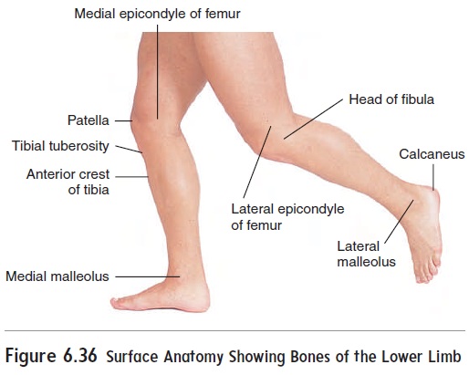
The thigh is the region between the hip and the knee (figure 6.33a). It contains a single bone called the femur. The head of the femur articulates with the acetabulum of the coxal bone. At the distal end of the femur, the condyles articulate with the tibia. Epicondyles, located medial and lateral to the condyles, are points of ligament attachment. The femur can be distinguished from the humerus by its long neck, located between the head and the trochanters
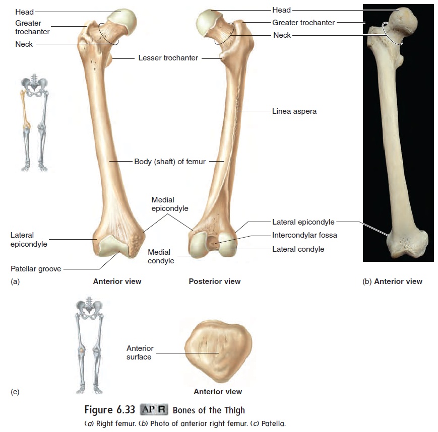
(trō′\kan-terz). A “broken hip” is usually a break of the femoral neck. A broken hip is difficult to repair and often requires pinning to hold the femoral head to the shaft. A major complication can occur if the blood vessels between the femoral head and the acetabulum are damaged. If this occurs, the femoral head may degenerate from lack of nourishment. The trochanters are points of muscle attach-ment. Thepatella (pa-tel′\ă), or kneecap (figure 6.33c), is located within the major tendon of the anterior thigh muscles and enables the tendon to bend over the knee.
Leg
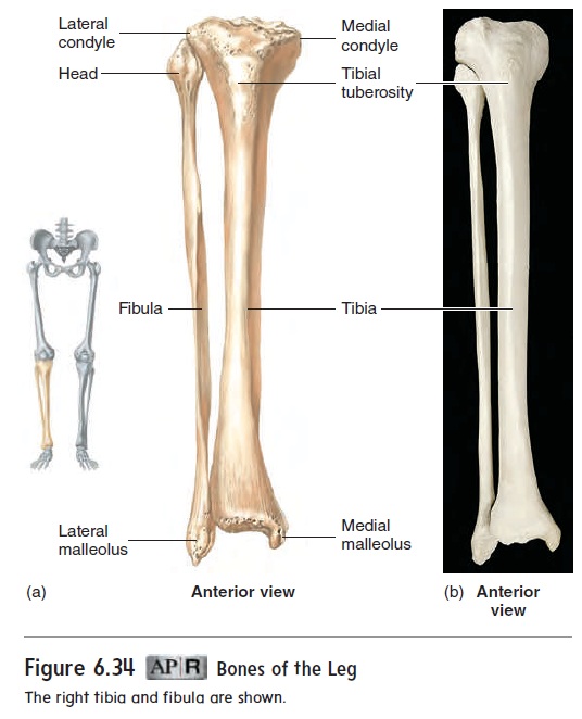
The leg is the region between the knee and the ankle (figure 6.34). It contains two bones, called the tibia (tib′\ē-ă; shinbone) and the fibula (fib′\ū-lă). The tibia is the larger of the two and is the majorweight-bearing bone of the leg. The rounded condyles of the femur rest on the flat condyles on the proximal end of the tibia. Just distal to the condyles of the tibia, on its anterior surface, is the tibialtuberosity, where the muscles of the anterior thigh attach. Thefibula does not articulate with the femur, but its head is attached to the proximal end of the tibia. The distal ends of the tibia and fibula form a partial socket that articulates with a bone of the ankle (the talus). A prominence can be seen on each side of the ankle (figure 6.34). These are the medial malleolus (mal-ē′\ō-lŭs) of the tibia and the lateral malleolus of the fibula.
Ankle
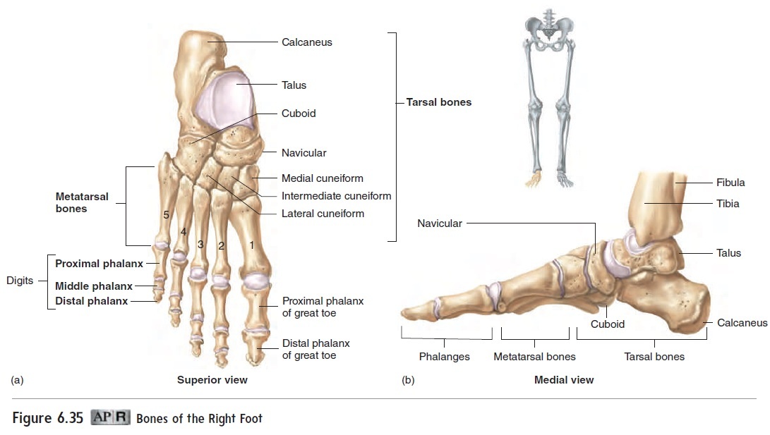
The ankle consists of seven tarsal (tar′\săl; the sole of the foot) bones (figure 6.35). The tarsal bones are the talus (tā′\lŭs; anklebone), calcaneus (kal-kā′\nē-ŭs; heel), cuboid (kū′\boyd), and navicular (nă-vik′\yū-lăr), and the medial, intermediate, and lateral cuneiforms (kū′\nē-i-fōrmz). The talus articulates with the tibiaand fibula to form the ankle joint, and the calcaneus forms the heel (figure 6.36). A mnemonic for the distal row is MILC—that is, Medial, Intermediate, and Lateral cuneiforms and the Cuboid. Amnemonic for the proximal three bones is No Thanks Cow—that is, Navicular, Talus, and Calcaneus.
Foot
The metatarsal (met′\ă-tar′\săl) bones and phalanges of the foot are arranged and numbered in a manner very similar to the meta-carpal bones and phalanges of the hand (see figure 6.35). The metatarsal bones are somewhat longer than the metacarpal bones, whereas the phalanges of the foot are considerably shorter than those of the hand.
There are three primary arches in the foot, formed by the positions of the tarsal bones and metatarsal bones, and held in place by ligaments. Two longitudinal arches extend from the heel to the ball of the foot, and a transverse arch extends across the foot. The arches function similarly to the springs of a car, allowing the foot to give and spring back.
Related Topics