Chapter: Human Neuroanatomy(Fundamental and Clinical): Gross Anatomy of the Cerebral Hemispheres
White Matter of Cerebral Hemispheres
WHITE MATTER OF CEREBRAL HEMISPHERES
Deep to the cerebral cortex the greater part of each cerebral hemisphere is occupied by nerve fibres that constitute the white matter. These fibres may be:
a. Association fibres that interconnect different regions of the cerebral cortex.
b. Projection fibres that connect the cerebral cortex with other masses of grey matter; and viceversa.
c. Commissural fibres that interconnect identical areas in the two hemispheres.
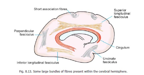
Association Fibres
These may be short and may connect adjoining gyri. Alternatively, they may be long and may connect distant parts of the cerebral cortex. Many of the association fibres form bundles that can be seen by gross dissection. Some of these are shown in Fig. 8.13. Some association fibres pass through commissures to connect dissimilar areas in the two cerebral hemispheres.
Projection Fibres
These fibres connect the cerebral cortex to centres in the brainstem and spinal cord, in both directions. Fibres to the cortex are often referred to as corticopetal fibres, while those going away from the cortex are referred to as corticofugal fibres. Fibres connecting the cortex with the thalamus, the hypothalamus and the basal ganglia, are also projection fibres. Many of the major projection fibres pass through the internal capsule, which is considered below.
The Internal Capsule
We have seen that a large number of nerve fibres interconnect the cerebral cortex with centres in the brainstem and spinal cord, and with the thalamus. Most of these fibres pass through the interval between the thalamus and caudate nucleus medially, and the lentiform nucleus laterally. This region
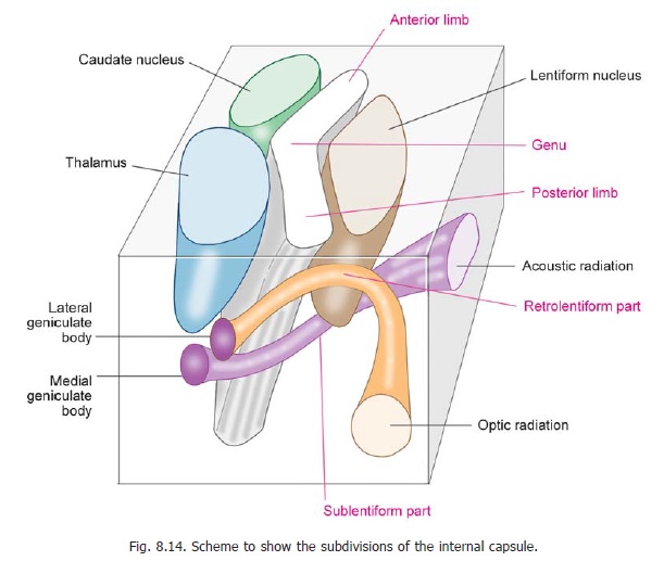
is called the internal capsule. Above, the internal capsule is continuous with the corona radiata; and, below, with the crus cerebri (of the midbrain).
The internal capsule may be divided into the following parts (Fig. 8.14).
1. The anterior limb lies between the caudate nucleus medially, and the anterior part of the lentiform nucleus laterally.
2. The posterior limb lies between the thalamus medially, and the posterior part of the lentiform nucleus on the lateral side.
3. In transverse sections through the cerebral hemisphere the anterior and posterior limbs of the internal capsule are seen to meet at an angle open outwards. This angle is called the genu (genu = bend).
4. Some fibres of the internal capsule lie behind the posterior end of the lentiform nucleus. They constitute its retrolentiform part.
5. Some other fibres pass below the lentiform nucleus (and not medial to it). These fibres constitute the sublentiform part of the internal capsule.
Corpus Callosum
The corpus callosum is made up of a large mass of nerve fibres that connects the two cerebral hemispheres (Fig. 8.6). It is subdivided into a central part or trunk, an anterior end that is bent on itself to form the genu, and an enlarged posterior end called thesplenium. A thin lamina of nerve
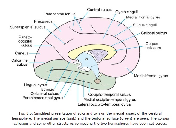
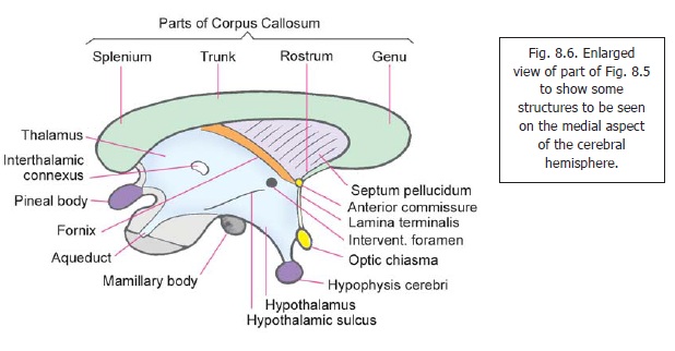
fibres connects the genu to the upper end of the lamina terminalis. These fibres form the rostrum of the corpus callosum. The corpus callosum is intimately related to the lateral ventricles. Its under surface gives attachment to the septum pellucidum (Figs. 8.6, 13.1).
The fibres of the corpus callosum interconnect the corresponding regions of almost all parts of the cerebral cortex of the two hemispheres. The fibres of the genu run forwards into the frontal lobes, the fibres of the two sides forming a fork-like structure called the forceps minor. Many fibres of the splenium run backwards into the occipital lobe to form a similar structure called the forceps major. (Each half of the forceps major bulges into the posterior horn of the corresponding lateral ventricle, forming the bulb of the posterior horn).
The fibres of the trunk of the corpus callosum (and some from the splenium) run laterally and as they do so they intersect the fibres of the corona radiata. As they pass laterally, some fibres of the trunk and of the splenium of the corpus callosum, form a flattened band called the tapetum. The tapetum is closely related to the posterior and inferior horns of the lateral ventricle.
As mentioned above all fibres passing through the corpus callosum are not strictly commissural. Some fibres that interconnect dissimilar areas in the two hemispheres are really association fibres.
Blood Supply of the Brain
A consolidated account of the blood supply of all parts of the brain.
Related Topics