Chapter: Basic & Clinical Pharmacology : Introduction to the Pharmacology of Central Nervous System (CNS) Drugs
The Synapse & Synaptic Potentials
THE SYNAPSE & SYNAPTIC
POTENTIALS
The
communication between neurons in the CNS occurs through chemical synapses in
the majority of cases. (A few instances of electrical coupling between neurons
have been documented, and such coupling may play a role in synchronizing
neuronal dis-charge. However, it is unlikely that these electrical synapses are
an important site of drug action.) The events involved in synaptic transmission
can be summarized as follows.
An action potential in the presynaptic fiber propagates into the synaptic terminal and activates voltage-sensitive calcium channels in the membrane of the terminal (see Figure 6–3). The calcium chan-nels responsible for the release of transmitter are generally resistant to the calcium channel-blocking agents discussed earily (verapamil, etc) but are sensitive to blockade by certain marine toxins and metal ions (see Tables 21–1 and 12–4). Calcium flows into the terminal, and the increase in intraterminal calcium concentration promotes the fusion of synaptic vesicles with the presynaptic mem-brane. The transmitter contained in the vesicles is released into the synaptic cleft and diffuses to the receptors on the postsynaptic mem-brane. Binding of the transmitter to its receptor causes a brief change in membrane conductance (permeability to ions) of the postsynaptic cell. The time delay from the arrival of the presynaptic action poten-tial to the onset of the postsynaptic response is approximately 0.5 ms. Most of this delay is consumed by the release process, par-ticularly the time required for calcium channels to open.
The
first systematic analysis of synaptic potentials in the CNS was in the early
1950s by Eccles and associates, who recorded intra-cellularly from spinal motor
neurons. When a microelectrode enters a cell, there is a sudden change in the
potential recorded by the electrode, which is typically about –70 mV (Figure
21–3). This is the resting membrane potential of the neuron. Two types of
pathways— excitatory and inhibitory—impinge on the motor neuron.
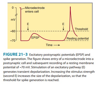
When an excitatory pathway is stimulated, a small depolariza-tion or excitatory postsynaptic potential (EPSP) is recorded. This potential is due to the excitatory transmitter acting on an ionotropic receptor, causing an increase in cation permeability. Changing the stimulus intensity to the pathway, and therefore the number of presynaptic fibers activated, results in a graded change in the size of the depolarization. When a sufficient number of excitatory fibers are activated, the excitatory postsynaptic potential depolarizes the postsynaptic cell to threshold, and an all-or-none action potential is generated.
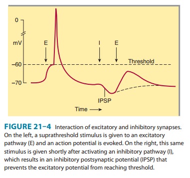
When
an inhibitory pathway is stimulated, the postsynaptic membrane is
hyperpolarized owing to the selective opening of chloride channels, producing
an inhibitory postsynaptic poten-tial
(IPSP) (Figure 21–4). However, because the equilibriumpotential for
chloride is only slightly more negative than the rest-ing potential (∼ –65 mV), the hyperpolarization is small and
contributes only modestly to the inhibitory action. The opening of the chloride
channel during the inhibitory postsynaptic poten-tial makes the neuron “leaky”
so that changes in membrane poten-tial are more difficult to achieve. This
shunting effect decreases the change in membrane potential during the
excitatory postsynaptic potential. As a result, an excitatory postsynaptic
potential that evoked an action potential under resting conditions fails to
evoke an action potential during the inhibitory postsynaptic potential (Figure
21–4). A second type of inhibition is presynaptic
inhibi-tion. It was first described for sensory fibers entering the
spinalcord, where excitatory synaptic terminals receive synapses called
axoaxonic synapses (described later). When activated, axoaxonic synapses reduce
the amount of transmitter released from the ter-minals of sensory fibers. It is
interesting that presynaptic inhibi-tory receptors are present on almost all
presynaptic terminals in the brain even though axoaxonic synapses appear to be
restricted to the spinal cord. In the brain, transmitter spills over to
neighbor-ing synapses to activate the presynaptic receptors.
Related Topics