Chapter: Medical Physiology: Pulmonary Circulation, Pulmonary Edema, Pleural Fluid
Physiologic Anatomy of the Pulmonary Circulatory System
Physiologic Anatomy of the Pulmonary Circulatory System
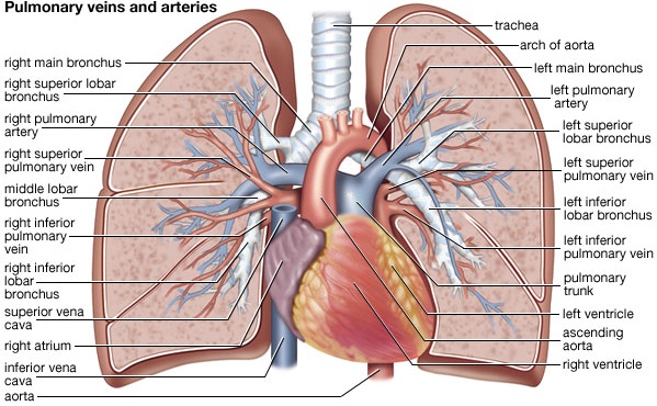
Pulmonary Vessels. The pulmonary artery extends only 5 centimeters beyond the apexof the right ventricle and then divides into right and left main branches that supply blood to the two respective lungs.
The pulmonary artery is thin, with a wall thickness one third that of the aorta. The pulmonary arterial branches are very short, and all the pulmonary arteries, even the smaller arteries and arterioles, have larger diameters than their counterpart sys-temic arteries. This, combined with the fact that the vessels are thin and distensible, gives the pulmonary arterial tree a large compliance, averaging almost 7 ml/mm Hg, which is similar to that of the entire systemic arterial tree. This large compliance allows the pulmonary arteries to accommodate the stroke volume output of the right ventricle.
The pulmonary veins, like the pulmonary arteries, are also short. They immedi-ately empty their effluent blood into the left atrium, to be pumped by the left heart through the systemic circulation.

Bronchial Vessels. Blood also flows to the lungs through small bronchial arteries thatoriginate from the systemic circulation, amounting to about 1 to 2 per cent of the total cardiac output. This bronchial arterial blood is oxygenated blood, in contrast to the partially deoxygenated blood in the pulmonary arteries. It supplies the sup-porting tissues of the lungs, including the connective tissue, septa, and large and small bronchi. After this bronchial and arterial blood has passed through the sup-porting tissues, it empties into the pulmonary veins and enters the left atrium, rather than passing back to the right atrium. Therefore, the flow into the left atrium and the left ventricular output are about 1 to 2 per cent greater than the right ventric-ular output.
Lymphatics. Lymph vessels are present in all the supportive tissues of the lung, begin-ning in the connective tissue spaces that surround the terminal bronchioles, cours-ing to the hilum of the lung, and thence mainly into the right thoracic lymph duct. Particulate matter entering the alveoli is partly removed by way of these channels, and plasma protein leaking from the lung capillaries is also removed from the lung tissues, thereby helping to prevent pulmonary edema.
Related Topics