Chapter: Ophthalmology: Vitreous Body
Persistent Fetal Vasculature (Developmental Anomalies)
Abnormal Changes in the Vitreous Body
Persistent Fetal Vasculature (Developmental Anomalies)
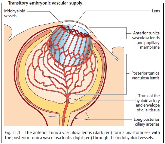
The embryonic vascular system in the vitreous
body and lens normally disap-pears completely, leaving only the hyaloid canal. Persistence of the
vascular system is referred to as persistent fetal vasculature. The following section describes the varying
degrees of severity of this syndrome as they relate to the vitreous body.
Persistence of the anterior tunica vasculosa lentis leads to a persistent
pupillary membrane.
Mittendorf’s Dot
Mittendorf’s dot is a small visually asymptomatic opacity in the
posterior lens capsule located approximately 0.5 mm medial to the center. This
is the site where the hyaloid artery enters the embryonic lens. This harmless
change occurs in up to 2% of the total population. Normal lens fiber
development can be disturbed where large portions of the hyaloid arterial
system remain, although this occurs very rarely. These patients develop
posterior polar cata-racts.
Bergmeister’s Papilla
Persistent Hyaloid Artery
Isolated persistence of the hyaloid artery is rare. Usually this phenomenon
isaccompanied by persistence of the hyperplastic primary vitreous (see next
section). A persistent hyaloid artery will appear as a whitish cord in the
hyaloid canal proceeding from the optic disk and extending to the posterior
capsule of the lens. Isolated persistence of the hyaloid artery is asymptomatic
and does not require treatment.
Persistent Hyperplastic Primary Vitreous (PHPV)
Definition
Persistence of the embryonic primary vitreous
(hyaloid arterial system includ-ing the posterior tunica vasculosa lentis).
Epidemiology:
This developmental anomaly is also very rare.
Symptoms and findings:
Usually the disorder isunilateral.
Anterior variant of PHPV.
With thismore frequentvariant,
awhite pupil(leukocoria or amaurotic
cat’s eye) typically will be discovered shortly after birth. This is caused by
the whitish plate of connective tissue posterior to the lens. Depending on the
severity, it will be accompanied by more or less severe changes in the lens
leading to more or less severely impaired vision. In extreme cases, the lens
resembles an opacified membrane (membranous
cat-aract). In rare cases, fatty
tissue will develop (lipomatous
pseudophakia), andeven more rarely cartilage
will develop in the lens. Retrolenticular scarring draws the ciliary processes
toward the center, and they will be visible in the pupil. Growth of the eye is
retarded. This results in microphthalmos
unless drainage of the aqueous humor is also impaired, in which case buphthalmos(hydrophthalmos) will be
present.
Posterior variant of PHPV.
Retinal detachment andretinal
dysplasiacan occurwhere primarily posterior embryonic structures persist.
The whitish plate of connective tissue will only be visible where anterior
changes associated with persistent hyperplastic primary vitreous are also
present. The reduction in visual acuity will vary depending on the severity of
the retinal changes.
Diagnostic considerations:
A definitive
diagnosis is usually possible on thebasis of the characteristic clinical
picture (see symptoms and findings) and additional ultrasound studies (when the
posterior segment is obscured by lens opacities).
Differential diagnosis:
Other causes of leukocoria (Table 11.1) should beexcluded. Retinoblastoma, the most important
differential diagnosis, can usually be excluded on the basis of ultrasound or
CT studies. In the presence of a retinoblastoma, these studies will reveal an
intraocular mass with calcifi-cations. In contrast to PHPV, microphthalmos will not be present.
Leukocoria should be regarded as a
retinoblastoma until proven other-wise.
Treatment:
The disorder is not usually treated as neither
conservative ther-apy nor surgery can improve visual acuity. Surgery is
indicated only where complications such as progressive collapse of the anterior
chamber, second-ary increase in intraocular pressure, vitreous hemorrhage, and
retinal detach-ment are present or imminent. The only goal is to save the eye
and maintain existing visual acuity.
Clinical course and prognosis:
The clinical course and prognosis depend pri-marily on the
severity of the disorder. However, adequate surgical interven-tion can often
save the eye and stabilize visual acuity even if at a very low level.
Related Topics