Chapter: Medical Physiology: Pulmonary Ventilation
Muscles That Cause Lung Expansion and Contraction
Muscles That Cause Lung Expansion and Contraction
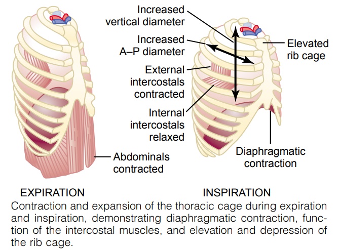
The lungs can be expanded and contracted in two ways: (1) by downward and upward movement of the diaphragm to lengthen or shorten the chest cavity, and (2) by elevation and depression of the ribs to increase and decrease the anteroposterior diameter of the chest cavity. Figure 37–1 shows these two methods.
Normal quiet breathing is accomplished almost entirely by the first method, that is, by movement of the diaphragm. During inspiration, contraction of the diaphragm pulls the lower surfaces of the lungs downward. Then, during expi-ration, the diaphragm simply relaxes, and the elastic recoil of the lungs, chest wall, and abdominal structures compresses the lungs and expels the air. During heavy breathing, however, the elastic forces are not powerful enough to cause the necessary rapid expiration, so that extra force is achieved mainly by con-traction of the abdominal muscles, which pushes the abdominal contents upward against the bottom of the diaphragm, thereby compressing the lungs.
The second method for expanding the lungs is to raise the rib cage. This expands the lungs because, in the natural resting position, the ribs slant down-ward, as shown on the left side of Figure 37–1, thus allowing the sternum to fall backward toward the vertebral column. But when the rib cage is elevated, the ribs project almost directly forward, so that the sternum also moves forward, away from the spine, making the anteroposterior thickness of the chest about 20 per cent greater during maximum inspiration than during expiration. There-fore, all the muscles that elevate the chest cage are classified as muscles of inspi-ration, and those muscles that depress the chest cage are classified as muscles of expiration. The most important muscles that raise the rib cage are the exter-nal intercostals, but others that help are the (1) sternocleidomastoid muscles,which lift upward on the sternum; (2) anterior serrati, which lift many of the ribs; and (3) scaleni, which lift the first two ribs.
The muscles that pull the rib cage downward during expiration are mainly the (1) abdominal recti, which have the powerful effect of pulling downward on the lower ribs at the same time that they and other abdominal muscles also com-press the abdominal contents upward against the diaphragm, and (2) internalintercostals.
Figure 37–1 also shows the mechanism by which the external and internal intercostals act to cause inspira-tion and expiration. To the left, the ribs during expiration are angled downward, and the external intercostals are elongated forward and downward. As they contract, they pull the upper ribs forward in rela-tion to the lower ribs, and this causes leverage on the ribs to raise them upward, thereby causing inspiration. The internal intercostals function exactly in the oppo-site manner, functioning as expiratory muscles because they angle between the ribs in the opposite direction and cause opposite leverage.
Related Topics