Chapter: Medical Physiology: Thyroid Metabolic Hormones
Diseases of the Thyroid
Diseases of the Thyroid
Hyperthyroidism
Most effects of hyperthyroidism are obvious from the preceding discussion of the various physiologic effects of thyroid hormone. However, some specific effects should be mentioned in connection especially with the development, diagnosis, and treatment of hyperthy-roidism.
Causes of Hyperthyroidism (Toxic Goiter, Thyrotoxicosis, Graves’ Disease). In most patients with hyperthyroidism, thethyroid gland is increased to two to three times normal size, with tremendous hyperplasia and infolding of the follicular cell lining into the follicles, so that the number of cells is increased greatly. Also, each cell increases its rate of secretion severalfold; radioactive iodine uptake studies indicate that some of these hyper-plastic glands secrete thyroid hormone at rates 5 to 15 times normal.
The changes in the thyroid gland in most instances are similar to those caused by excessive TSH. However, plasma TSH concentrations are less than normal rather than enhanced in almost all patients and often are essentially zero. However, other substances that have actions similar to those of TSH are found in the blood of almost all these patients. These substances are immunoglobulin antibodies that bind with the same membrane receptors that bind TSH. They induce continual activation of the cAMP system of the cells, with resultant development of hyperthyroidism. These antibodies are called thyroid-stimulating immunoglobu-lin and are designated TSI. They have a prolongedstimulating effect on the thyroid gland, lasting for as long as 12 hours, in contrast to a little over 1 hour for TSH. The high level of thyroid hormone secretion caused by TSI in turn suppresses anterior pituitary for-mation of TSH.
The antibodies that cause hyperthyroidism almost certainly occur as the result of autoimmunity that has developed against thyroid tissue. Presumably, at some time in the history of the person, an excess of thyroid cell antigens was released from the thyroid cells, and this has resulted in the formation of antibodies against the thyroid gland itself.
Thyroid Adenoma. Hyperthyroidism occasionally resultsfrom a localized adenoma (a tumor) that develops in the thyroid tissue and secretes large quantities of thyroid hormone. This is different from the more usual type of hyperthyroidism, in that it usually is not associated with evidence of any autoimmune disease. An interesting effect of the adenoma is that as long as it continues to secrete large quantities of thyroid hormone, secretory function in the remainder of the thyroid gland is almost totally inhibited because the thyroid hormone from the adenoma depresses the production of TSH by the pitu-itary gland.
Symptoms of Hyperthyroidism
The symptoms of hyperthyroidism are obvious from the preceding discussion of the physiology of the thyroid hormones: (1) a high state of excitability, (2) intolerance to heat, (3) increased sweating, (4) mild to extreme weight loss (sometimes as much as 100 pounds), (5) varying degrees of diarrhea, (6) muscle weakness, (7) nervousness or other psychic disorders, (8) extreme fatigue but inability to sleep, and (9) tremor of the hands.
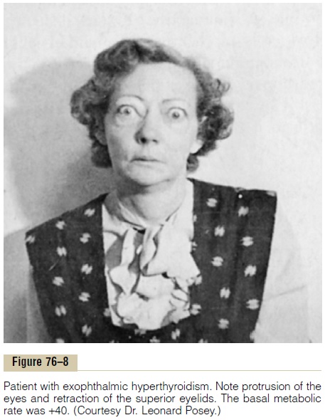
Exophthalmos. Most people with hyperthyroidismdevelop some degree of protrusion of the eyeballs, as shown in Figure 76–8. This condition is called exoph-thalmos. A major degree of exophthalmos occurs inabout one third of hyperthyroid patients, and the con-dition sometimes becomes so severe that the eyeball protrusion stretches the optic nerve enough to damage vision. Much more often, the eyes are damaged because the eyelids do not close completely when the person blinks or is asleep. As a result, the epithelial surfaces of the eyes become dry and irritated and often infected, resulting in ulceration of the cornea.
The cause of the protruding eyes is edematous swelling of the retro-orbital tissues and degenerative changes in the extraocular muscles. In most patients, immunoglobulins can be found in the blood that react with the eye muscles. Furthermore, the concentration of these immunoglobulins is usually highest in patients who have high concentrations of TSIs. Therefore, there is much reason to believe that exophthalmos, like hyper-thyroidism itself, is an autoimmune process. The exoph-thalmos usually is greatly ameliorated with treatment of the hyperthyroidism.
Diagnostic Tests for Hyperthyroidism. For the usual case ofhyperthyroidism, the most accurate diagnostic test is direct measurement of the concentration of “free” thy-roxine (and sometimes triiodothyronine) in the plasma, using appropriate radioimmunoassay procedures.
Other tests that are sometimes used are as follows:
1.The basal metabolic rate is usually increased to +30 to +60 in severe hyperthyroidism.
2.The concentration of TSH in the plasma is measured by radioimmunoassay. In the usual type of thyrotoxicosis, anterior pituitary secretion of TSH is so completely suppressed by thelarge amounts of circulating thyroxine and triiodothyronine that there is almost no plasma TSH.
3. The concentration of TSI is measured by radioimmunoassay. This is usually high in thyrotoxicosis but low in thyroid adenoma.
Physiology of Treatment in Hyperthyroidism. The most directtreatment for hyperthyroidism is surgical removal of most of the thyroid gland. In general, it is desirable to prepare the patient for surgical removal of the gland before the operation. This is done by administering propylthiouracil, usually for several weeks, until the basal metabolic rate of the patient has returned to normal. Then, administration of high concentrations of iodides for 1 to 2 weeks immediately before operation causes the gland itself to recede in size and its blood supply to diminish. By using these preoperative proce-dures, the operative mortality is less than 1 in 1000 in the better hospitals, whereas before development of modern procedures, operative mortality was 1 in 25.
Treatment of the Hyperplastic Thyroid Gland with Radioactive Iodine
Eighty to 90 per cent of an injected dose of iodide is absorbed by the hyperplastic, toxic thyroid gland within 1 day after injection. If this injected iodine is radio-active, it can destroy most of the secretory cells of the thyroid gland. Usually 5 millicuries of radioactive iodine is given to the patient, whose condition is reassessed several weeks later. If the patient is still hyperthyroid, additional doses are administered until normal thyroid status is reached.
Hypothyroidism
The effects of hypothyroidism, in general, are opposite to those of hyperthyroidism, but there are a few physiologic mechanisms peculiar to hypothyroidism. Hypothyroidism, like hyperthyroidism, probably is ini-tiated by autoimmunity against the thyroid gland, but immunity that destroys the gland rather than stimulates it. The thyroid glands of most of these patients first have autoimmune “thyroiditis,” which means thyroid inflam-mation.This causes progressive deterioration and finally fibrosis of the gland, with resultant diminished or absent secretion of thyroid hormone. Several other types of hypothyroidism also occur, often associated with devel-opment of enlarged thyroid glands, called thyroid goiter, as follows.
Endemic Colloid Goiter Caused by Dietary Iodide Deficiency. Theterm “goiter” means a greatly enlarged thyroid gland. As pointed out in the discussion of iodine metabolism, about 50 milligrams of iodine are required each year for the formation of adequate quantities of thyroid hormone. In certain areas of the world, notably in the Swiss Alps, the Andes, and the Great Lakes region of the United States, insufficient iodine is present in the soil for the foodstuffs to contain even this minute quan-tity. Therefore, in the days before iodized table salt, many people who lived in these areas developed extremely large thyroid glands, called endemic goiters.
The mechanism for development of large endemic goiters is the following: Lack of iodine prevents pro-duction of both thyroxine and triiodothyronine. As a result, no hormone is available to inhibit production of TSH by the anterior pituitary; this causes the pituitary to secrete excessively large quantities of TSH. The TSH then stimulates the thyroid cells to secrete tremendous amounts of thyroglobulin colloid into the follicles, and the gland grows larger and larger. But because of lack of iodine, thyroxine and triiodothyronine production does not occur in the thyroglobulin molecule and there-fore does not cause the normal suppression of TSH pro-duction by the anterior pituitary. The follicles become tremendous in size, and the thyroid gland may increase to 10 to 20 times normal size.
Idiopathic Nontoxic Colloid Goiter. Enlarged thyroid glandssimilar to those of endemic colloid goiter can also occur in people who do not have iodine deficiency. These goitrous glands may secrete normal quantities of thyroid hormones, but more frequently, the secretion of hormone is depressed, as in endemic colloid goiter.
The exact cause of the enlarged thyroid gland in patients with idiopathic colloid goiter is not known, but most of these patients show signs of mild thyroiditis; therefore, it has been suggested that the thyroiditis causes slight hypothyroidism, which then leads to increased TSH secretion and progressive growth of the noninflamed portions of the gland. This could explain why these glands usually are nodular, with some por-tions of the gland growing while other portions are being destroyed by thyroiditis.
In some persons with colloid goiter, the thyroid gland has an abnormality of the enzyme system required for formation of the thyroid hormones. Among the abnor-malities often encountered are the following:
1.Deficient iodide-trapping mechanism, in whichiodine is not pumped adequately into the thyroid cells
2.Deficient peroxidase system, in which the iodidesare not oxidized to the iodine state
3.Deficient coupling of iodinated tyrosines in the thyroglobulin molecule, so that the final thyroidhormones cannot be formed
4.Deficiency of the deiodinase enzyme, whichprevents recovery of iodine from the iodinated tyrosines that are not coupled to form the thyroid hormones (this is about two thirds of the iodine),
thus leading to iodine deficiency
Finally, some foods contain goitrogenic substances that have a propylthiouracil-type of antithyroid activity, thus also leading to TSH-stimulated enlargement of the thyroid gland. Such goitrogenic substances are found especially in some varieties of turnips and cabbages.
Physiologic Characteristics of Hypothyroidism. Whetherhypothyroidism is due to thyroiditis, endemic colloid goiter, idiopathic colloid goiter, destruction of the thyroid gland by irradiation, or surgical removal of the thyroid gland, the physiologic effects are the same. They include fatigue and extreme somnolence with sleeping up to 12 to 14 hours a day, extreme muscular sluggish-ness, slowed heart rate, decreased cardiac output, decreased blood volume, sometimes increased body weight, constipation, mental sluggishness, failure of many trophic functions in the body evidenced by depressed growth of hair and scaliness of the skin, development of a froglike husky voice, and, in severe cases, development of an edematous appearance throughout the body called myxedema.
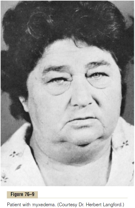
Myxedema. Myxedemadevelops in the patient withalmost total lack of thyroid hormone function. Figure 76–9 shows such a patient, demonstrating bagginess under the eyes and swelling of the face. In this condition, for reasons not explained, greatly increased quantities of hyaluronic acid and chondroitin sulfate bound with protein form excessive tissue gel in the interstitial spaces, and this causes the total quantity of interstitial fluid to increase. Because of the gel nature of the excess fluid, it is mainly immobile, and the edema is the nonpitting type.
Atherosclerosis in Hypothyroidism. As pointed out earlier,lack of thyroid hormone increases the quantity of blood cholesterol because of altered fat and cholesterol metabolism and diminished liver excretion of cholesterol in the bile. The increase in blood cholesterol is usually associated with increased atherosclerosis. Therefore, many hypothyroid patients, particularly those with myxedema, develop atherosclerosis, which in turn results in peripheral vascular disease, deafness, and coronary artery disease with consequent early death.
Diagnostic Tests in Hypothyroidism. The tests alreadydescribed for diagnosis of hyperthyroidism give oppo-site results in hypothyroidism. The free thyroxine in the blood is low. The basal metabolic rate in myxedema ranges between -30 and -50. And the secretion of TSH by the anterior pituitary when a test dose of TRH is administered is usually greatly increased (except in those rare instances of hypothyroidism caused by depressed response of the pituitary gland to TRH).
Treatment of Hypothyroidism. Figure 76–4 shows the effectof thyroxine on the basal metabolic rate, demonstrating that the hormone normally has a duration of action of more than 1 month. Consequently, it is easy to maintain a steady level of thyroid hormone activity in the body by daily oral ingestion of a tablet or more containing thyroxine. Furthermore, proper treatment of the hypothyroid patient results in such complete normality that formerly myxedematous patients have lived into their 90s after treatment for more than 50 years.
Cretinism
Cretinism is caused by extreme hypothyroidism during fetal life, infancy, or childhood. This condition is char-acterized especially by failure of body growth and by mental retardation. It results from congenital lack of a thyroid gland (congenital cretinism), from failure of the thyroid gland to produce thyroid hormone because of a genetic defect of the gland, or from iodine lack in the diet (endemic cretinism). The severity of endemic cre-tinism varies greatly, depending on the amount of iodine in the diet, and whole populaces of an endemic geo-graphic iodine-deficient soil area have been known to have cretinoid tendencies.
A neonate without a thyroid gland may have normal appearance and function because it was supplied with some (but usually not enough) thyroid hormone by the mother while in utero, but a few weeks after birth, the neonate’s movements become sluggish and both physi-cal and mental growth begin to be greatly retarded. Treatment of the neonate with cretinism at any time with adequate iodine or thyroxine usually causes normal return of physical growth, but unless the cre-tinism is treated within a few weeks after birth, mental growth remains permanently retarded. This results from retardation of the growth, branching, and myelination of the neuronal cells of the central nervous system at this critical time in the normal development of the mental powers.
Skeletal growth in the child with cretinism is charac-teristically more inhibited than is soft tissue growth. As a result of this disproportionate rate of growth, the soft tissues are likely to enlarge excessively, giving the child with cretinism an obese, stocky, and short appearance. Occasionally the tongue becomes so large in relation to the skeletal growth that it obstructs swallowing and breathing, inducing a characteristic guttural breathing that sometimes chokes the child.
Related Topics