Chapter: Essentials of Anatomy and Physiology: Blood Vessels and Circulation
Blood Vessels of the Systemic Circulation: Veins
BLOOD VESSELS OF THE SYSTEMIC CIRCULATION: VEINS
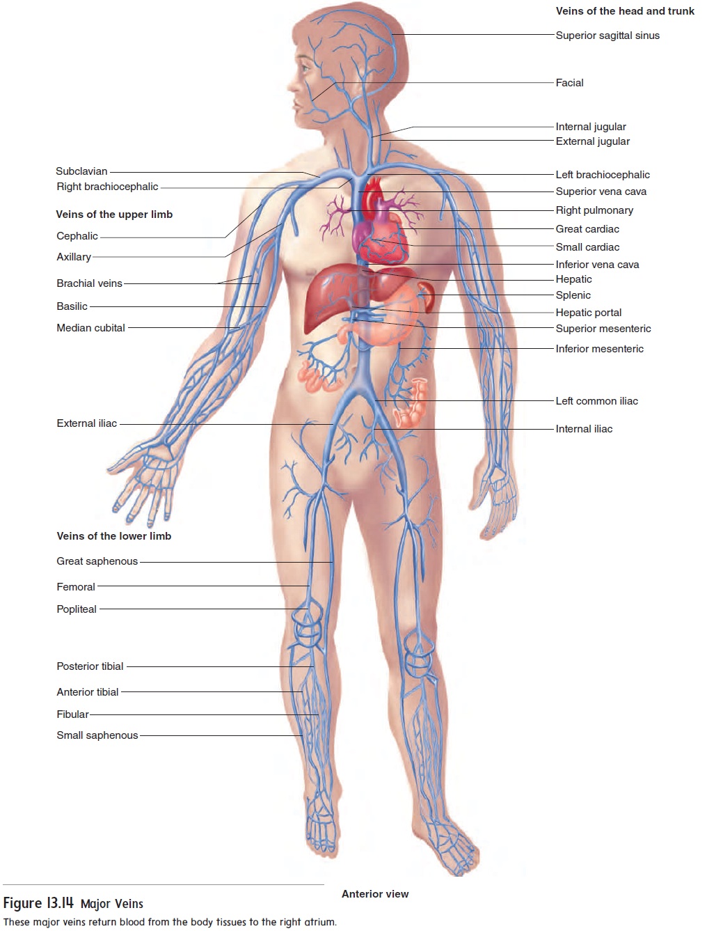
The oxygen-poor blood from the tissues of the body returns to the heart through veins. The superior vena cava (vē′ nă kā′ vă) returns blood from the head, neck, thorax, and upper limbs to the right atri-um of the heart, and the inferior vena cava returns blood from the abdomen, pelvis, and lower limbs to the right atrium (figure 13.14).
Veins of the head and neck
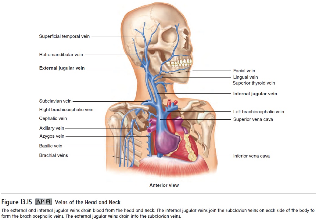
The two pairs of major veins that drain blood from the head and neck are the external and internal jugular (jŭg′ ū-lar) veins (fig-ure 13.15). The external jugular veins are the more superficial of the two sets. They drain blood from the posterior head and neck, emptying primarily into the subclavian veins. The internal jugular veins are much larger and deeper. They drain blood from the brain and the anterior head, face, and neck. The internal jugular veins join the subclavian veins on each side of the body to form the brachiocephalic veins. The brachiocephalic veins join to formthe superior vena cava.
Veins of the upper limbs
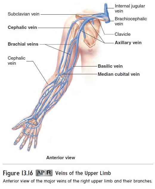
The veins of the upper limbs (figure 13.16) can be divided into deep and superficial groups. The deep veins, which drain the deep structures of the upper limbs, follow the same course as the arteries and are named for their respective arteries. The only noteworthy deep veins are the brachial veins, which accompany the brachial artery and empty into the axillary vein. The superficial veins drain the superficial structures of the upper limbs and then empty into the deep veins. The cephalic (sĕ′ fal′ ik; toward the head) vein, which empties into the axillary vein, and the basilic (ba-sil′ ik) vein, which becomes the axillary vein, are the major superficialveins. Many of their tributaries in the forearm and hand can be seen through the skin. The median cubital (kū′ bi-tal) vein usually connects the cephalic vein or its tributaries with the basilic vein. Although this vein varies in size among people, it is usually quite prominent on the anterior surface of the upper limb at the level of the elbow, an area called the cubital fossa,and is often used as a site for drawing blood.
Veins of the thorax
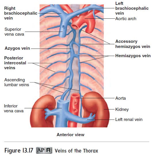
Three major veins return blood from the thorax to the superior vena cava: the right and left brachiocephalic veins and the azy-gos (az-ı̆′gos, az′i-gos) vein (figure 13.17). Blood drains from theanterior thoracic wall by way of the anterior intercostal veins. These veins empty into the internal thoracic veins, which empty into the brachiocephalic veins. Blood from the posterior thoracic wall is collected byposterior intercostal veins that drain into the azygos vein on the right and the hemiazygos veinor the acces-sory hemiazygos vein on the left. The hemiazygos and accessoryhemiazygos veins empty into the azygos vein, which drains into the superior vena cava (see figure 13.15).
Veins of the Abdomen and Pelvis
Figure 13.18 provides an overview of the major veins of the abdo-men and pelvis. It may help to refer to this figure as you read the following discussion. Blood from the posterior abdominal wall drains through ascending lumbar veins into the azygos vein. Blood from the rest of the abdomen and from the pelvis and lower limbs returns to the heart through the inferior vena cava. The gonads (testes or ovaries), kidneys, adrenal glands, and liver are the only abdominal organs outside the pelvis from which blood drains directly into the inferior vena cava. The internal iliac veins drain the pelvis and join theexternal iliac veins from the lower limbs to form the common iliac veins. The common iliac veins combine to form the inferior vena cava (see figure 13.14).
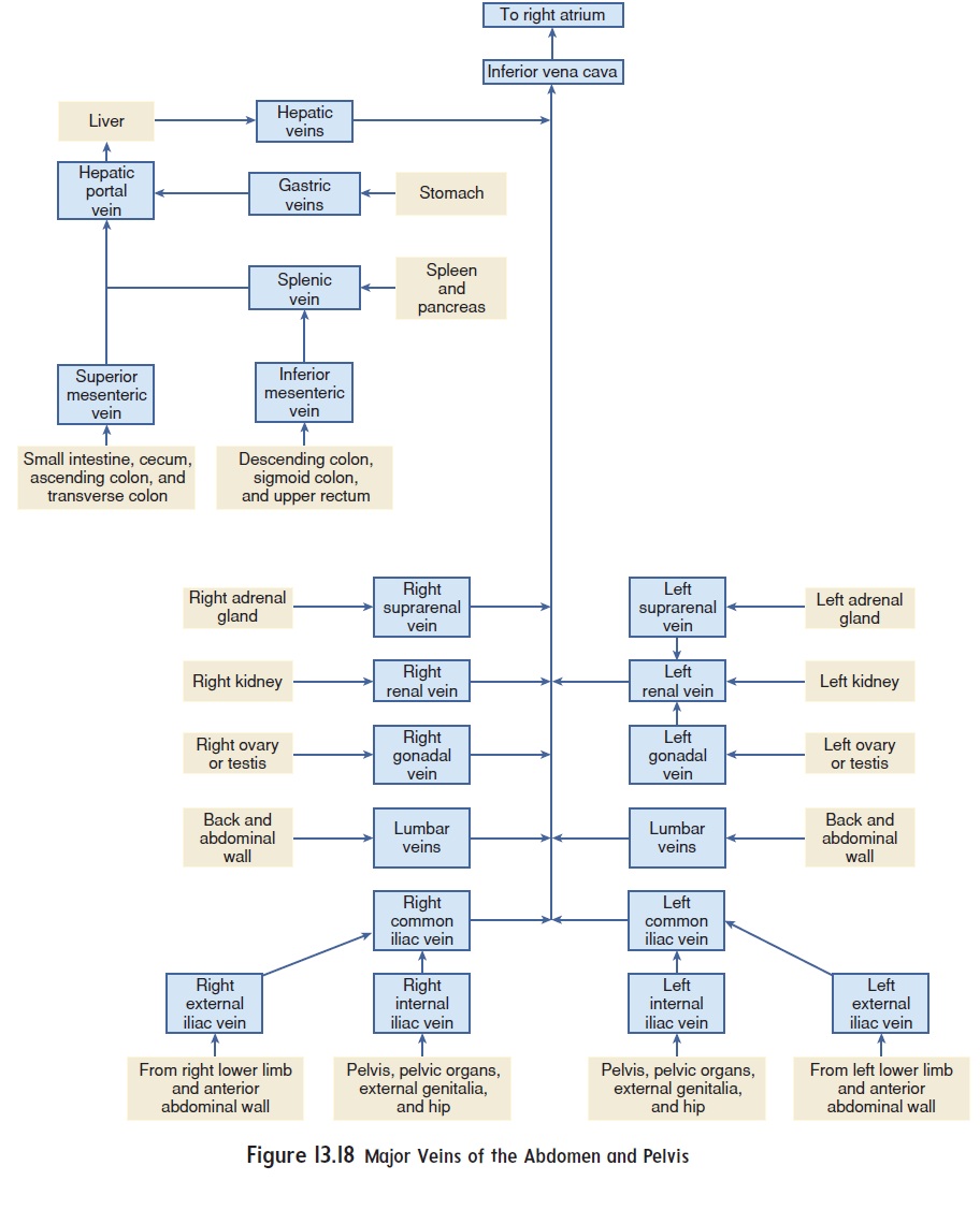
Blood from the capillaries within most of the abdominal viscera, such as the stomach, intestines, pancreas, and spleen, drains through a specialized system of blood vessels to the liver. The liver is a major processing center for substances absorbed by the intestinal tract. A portal (pōr′ tăl) system is a vascular system that begins and ends with capillary beds and has no pumping mechanism, such as the heart, in between. The hepatic (he-pa′ tik) portal system (figure 13.19) begins with capillariesin the viscera and ends with capillaries in the liver. The major tributaries of the hepatic portal system are the splenic(splen′ ik) vein and the superior mesenteric vein. The inferior mesenteric vein empties into the splenic vein. The splenic vein carries bloodfrom the spleen and pancreas. The superior and inferior mesen-teric veins carry blood from the intestines. The splenic vein and the superior mesenteric vein join to form the hepatic portalvein, which enters the liver.
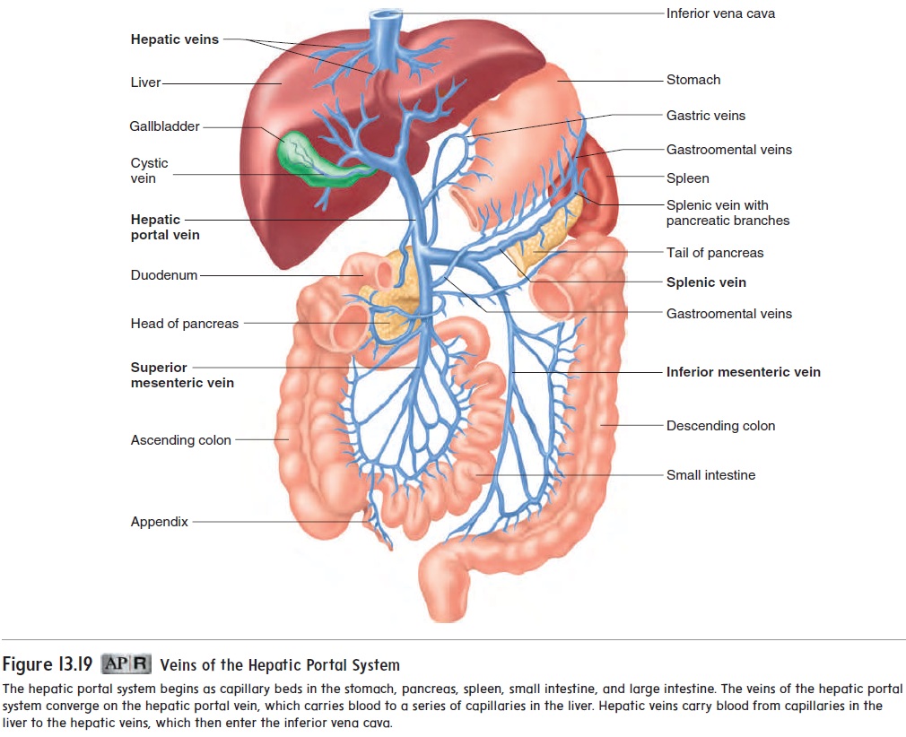
Blood from the liver flows into hepatic veins, which join the inferior vena cava. Blood entering the liver through the hepatic portal vein is rich in nutrients collected from the intestines, but it may also contain a number of toxic substances that are potentially harmful to body tissues. Within the liver, nutrients are taken up and stored or modified, so that they can be used by other cells of the body. Also within the liver, toxic substances are converted to nontoxic substances. These substances can be removed from the blood or carried by the blood to the kidneys for excretion.
Other veins of the abdomen and pelvis include the renal veins, the suprarenal veins, and the gonadal veins. The renal veins drain the kidneys, and the suprarenal veins drain the adrenal glands. The testicular veins drain the testes in males; the ovarian veins drain the ovaries in females.
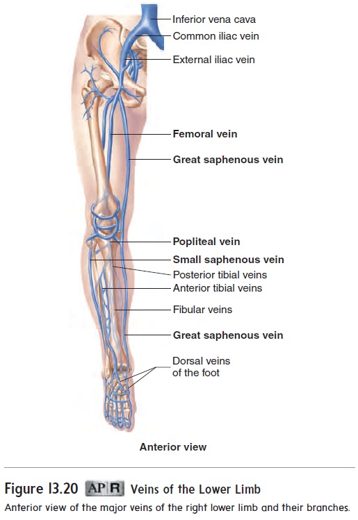
Veins of the lower limbs
The veins of the lower limbs (figure 13.20), like those of the upper limbs, consist of deep and superficial groups. The deep veins fol-low the same path as the arteries and are named for the arteries they accompany. The superficial veins consist of the great and small saphenous (să-fē′ nŭs, sa′ fĕ-nŭs) veins. The great saphe-nous vein originates over the dorsal and medial side of the footand ascends along the medial side of the leg and thigh to empty into the femoral vein. The small saphenous vein begins over the lateral side of the foot and joins the popliteal vein, which becomes the femoral vein. The femoral vein empties into the external iliac vein.
Related Topics