Chapter: Medical Physiology: The Eye: II. Receptor and Neural Function of the Retina
Anatomy and Function of the Structural Elements of the Retina
Anatomy and Function of the Structural Elements of the Retina
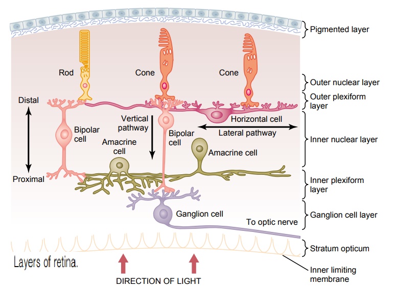
Layers of the Retina. Figure 50–1 shows the functional components of the retina whichare arranged in layers from the outside to the inside as follows: (1) pigmented layer, (2) layer of rods and cones projecting to the pigment, (3) outer nuclear layer con-taining the cell bodies of the rods and cones, (4) outer plexiform layer, (5) inner nuclear layer, (6) inner plexiform layer, (7) ganglionic layer, (8) layer of optic nerve fibers, and (9) inner limiting membrane.
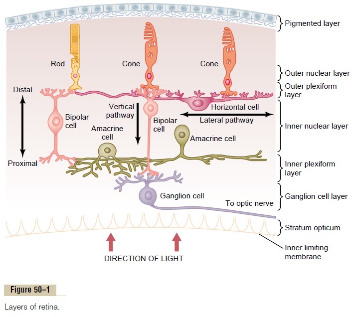
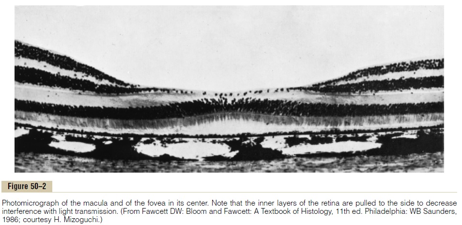
After light passes through the lens system of the eye and then through the vitre-ous humor, it enters the retina from the inside(see Figure 50–1); that is, it passes first through the ganglion cells and then through the plexiform and nuclear layers before it finally reaches the layer of rods and cones located all the way on the outer edge of the retina. This distance is a thickness of several hundred micrometers; visual acuity is decreased by this passage through such nonhomogeneous tissue. However, in the central foveal region of the retina, as discussed subsequently, the inside layers are pulled aside to decrease this loss of acuity.
Foveal Region of the Retina and Its Importance in Acute Vision. Thefoveais a minute areain the center of the retina, shown in Figure 50–2, occupying a total area a little more than 1 square millimeter; it is especially capable of acute and detailed vision. The central fovea, only 0.3 millimeter in diameter, is composed almost entirely of cones;these cones have a special structure that aids their detection of detail in the visual image. That is, the foveal cones have especially long and slender bodies, in con-tradistinction to the much fatter cones located more peripherally in the retina. Also, in the foveal region, the blood vessels, ganglion cells, inner nuclear layer of cells, and plexiform layers are all displaced to one side rather than resting directly on top of the cones. This allows light to pass unimpeded to the cones.
Rods and Cones. Figure 50–3 is a diagrammatic representation of the essential com-ponents of a photoreceptor (either a rod or a cone). As shown in Figure 50–4, the outer segment of the cone is conical in shape. In general, the rods are narrower and longer than the cones, but this is not always the case. In the peripheral portions of the retina, the rods are 2 to 5 micrometers in diameter, whereas the cones are 5 to 8 micrometers in diameter; in the central part of the retina, in the fovea, there are rods, and the cones are slender and have a diameter of only 1.5 micrometers.
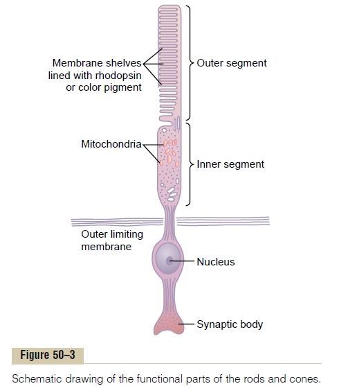
To the right in Figure 50–3 are labeled the major functional segments of either a rod or a cone: (1) the outer segment, (2) theinner segment, (3) the nucleus, and (4) the synaptic body. The light-sensitive photochemical is found in the outer segment. In the case of the rods, this is rhodopsin; in the cones, it is one of three “color” photochemicals, usually called simply color pigments, that function almost exactly the same as rhodopsin except for differences in spectral sensitivity.
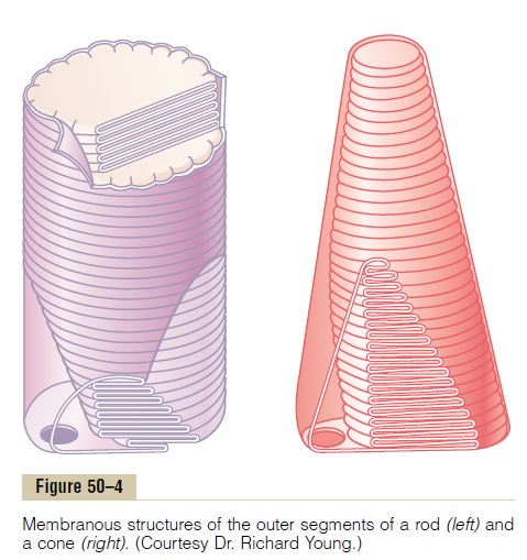
Note in the outer segments of the rods and cones in Figures 50–3 and 50–4 the large numbers of discs. Each of the discs is actually an infolded shelf of cell mem-brane. There are as many as 1000 discs in each rod or cone.
Both rhodopsin and the color pigments are conju-gated proteins. They are incorporated into the mem-branes of the discs in the form of transmembrane proteins. The concentrations of these photosensitive pigments in the discs are so great that the pigments themselves constitute about 40 per cent of the entire mass of the outer segment.
The inner segment of the rod or cone contains the usual cytoplasm with cytoplasmic organelles. Particu-larly important are the mitochondria; as explained later, these mitochondria play the important role of provid-ing energy for function of the photoreceptors.
The synaptic body is the portion of the rod or cone that connects with subsequent neuronal cells, the horizontal and bipolar cells, that represent the nextstages in the vision chain.
Pigment Layer of the Retina. The black pigmentmelanininthe pigment layer prevents light reflection throughout the globe of the eyeball; this is extremely important for clear vision. This pigment performs the same function in the eye as the black coloring inside the bellows of a camera. Without it, light rays would be reflected in all directions within the eyeball and would cause diffuse lighting of the retina rather than the normal contrast between dark and light spots required for formation of precise images.
The importance of melanin in the pigment layer is well illustrated by its absence in albinos, people who are hereditarily lacking in melanin pigment in all parts of their bodies. When an albino enters a bright room, light that impinges on the retina is reflected in all directions inside the eyeball by the unpigmented surfaces of the retina and by the underlying sclera, so that a single discrete spot of light that would normally excite only a few rods or cones is reflected everywhere and excites many receptors. Therefore, the visual acuity of albinos, even with the best optical correction, is seldom better than 20/100 to 20/200 rather than the normal 20/20 values.
The pigment layer also stores large quantities of vitamin A. This vitamin A is exchanged back and forththrough the cell membranes of the outer segments of the rods and cones, which themselves are embedded in the pigment. We show later that vitamin A is an impor-tant precursor of the photosensitive chemicals of the rods and cones.
Blood Supply of the Retina—The Central Retinal Artery and the Choroid. The nutrient blood supply for the internallayers of the retina is derived from the central retinal artery, which enters the eyeball through the center of the optic nerve and then divides to supply the entireinside retinal surface. Thus, the inner layers of the retinahave their own blood supply independent of the other structures of the eye.
However, the outermost layer of the retina is adherent to the choroid, which is also a highly vascular tissue lying between the retina and the sclera. The outer layers of the retina, especially the outer segments of the rods and cones, depend mainly on diffusion from the choroid blood vessels for their nutrition, especially for their oxygen.
Retinal Detachment. The neural retina occasionallydetaches from the pigment epithelium. In some instances,the cause of such detachment is injury to the eyeball that allows fluid or blood to collect between the neural retina and the pigment epithelium. Detachment is occa-sionally caused by contracture of fine collagenous fibrils in the vitreous humor, which pull areas of the retina toward the interior of the globe.
Partly because of diffusion across the detachment gap and partly because of the independent blood supply to the neural retina through the retinal artery, the detached retina can resist degeneration for days and can become functional again if it is surgically replaced in its normal relation with the pigment epithelium. If it is not replaced soon, however, the retina will be destroyed and will be unable to function even after surgical repair.
Related Topics