Chapter: Medical Physiology: Cardiac Arrhythmias and Their Electrocardiographic Interpretation
Abnormal Sinus Rhythms
Abnormal Sinus Rhythms
Tachycardia
The term “tachycardia” means fast heart rate, usually defined in an adult person as faster than 100 beats per minute. An electrocardiogram recorded from a patient with tachycardia is shown in Figure 13–1. This electrocardiogram is normal except that the heart rate, as determined from the time intervals between QRS complexes, is about 150 per minute instead of the normal 72 per minute.
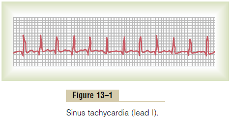
The general causes of tachycardia include increased body temperature, stimulationof the heart by the sympathetic nerves, or toxic conditions of the heart.
The heart rate increases about 10 beats per minute for each degree Fahrenheit (18 beats per degree Celsius) increase in body temperature, up to a body tempera-ture of about 105°F (40.5°C); beyond this, the heart rate may decrease because of progressive debility of the heart muscle as a result of the fever. Fever causes tachy-cardia because increased temperature increases the rate of metabolism of the sinus node, which in turn directly increases its excitability and rate of rhythm.
Many factors can cause the sympathetic nervous system to excite the heart, as we discuss at multiple points in this text. For instance, when a patient loses blood and passes into a state of shock or semishock, sympathetic reflex stimulation of the heart often increases the heart rate to 150 to 180 beats per minute.
Simple weakening of the myocardium usually increases the heart rate because the weakened heart does not pump blood into the arterial tree to a normal extent, and this elicits sympathetic reflexes to increase the heart rate.
Bradycardia
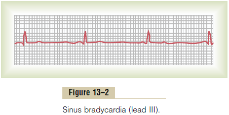
The term “bradycardia” means a slow heart rate, usually defined as fewer than 60 beats per minute. Bradycardia is shown by the electrocardiogram in Figure 13–2.
Bradycardia in Athletes. The athlete’s heart is larger and considerably stronger thanthat of a normal person, which allows the athlete’s heart to pump a large stroke volume output per beat even during periods of rest. When the athlete is at rest, excessive quantities of blood pumped into the arterial tree with each beat initiate feedback circulatory reflexes or other effects to cause bradycardia.
Vagal Stimulation as a Cause of Bradycardia. Any circulatoryreflex that stimulates the vagus nerves causes release of acetylcholine at the vagal endings in the heart, thus giving a parasympathetic effect. Perhaps the most strik-ing example of this occurs in patients with carotid sinussyndrome. In these patients, the pressure receptors(baroreceptors) in the carotid sinus region of the carotid artery walls are excessively sensitive. Therefore, even mild external pressure on the neck elicits a strong baroreceptor reflex, causing intense vagal-acetylcholine effects on the heart, including extreme bradycardia. Indeed, sometimes this reflex is so powerful that it actu-ally stops the heart for 5 to 10 seconds.
Sinus Arrhythmia
Figure 13–3 shows a cardiotachometer recording of the heart rate, at first during normal and then (in the second half of the record) during deep respiration. A cardiota-chometer is an instrument that records by the height ofsuccessive spikes the duration of the interval betweenthe successive QRS complexes in the electrocar-diogram. Note from this record that the heart rate increased and decreased no more than 5 per cent during
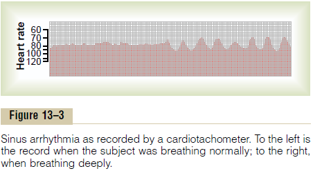
quiet respiration (left half of the record). Then, duringdeep respiration, the heart rate increased and decreasedwith each respiratory cycle by as much as 30 per cent.
Sinus arrhythmia can result from any one of many cir-culatory conditions that alter the strengths of the sym-pathetic and parasympathetic nerve signals to the heart sinus node. In the “respiratory” type of sinus arrhyth-mia, as shown in Figure 13–3, this results mainly from “spillover” of signals from the medullary respiratory center into the adjacent vasomotor center during inspi-ratory and expiratory cycles of respiration. The spillover signals cause alternate increase and decrease in the number of impulses transmitted through the sympa-thetic and vagus nerves to the heart.
Related Topics