Chapter: Ophthalmology: Visual Pathway
Visual Pathway: Examination Methods
Examination Methods
Visual field testing (perimetry):
This is the most important test for visualpathway lesions.
Because it permits one to diagnose the location of the lesion, it is also of
interest from a neurologic standpoint. The “visual
field” is defined as the field of perception
of the eye at rest with the gaze directed straight ahead. It includes all
points (objects and surfaces) in space that are simultaneously visible when the
eye focuses on one point.
The examination is performed on one eye at a time. The principle of the test is to have the patient focus on a central point in
the device while the eye is in a defined state of adaptation with controlled
ambient lighting (see below). Light markers appear in the hemisphere of the
device. The patient signals that he or she perceives the markers by pressing a
button that triggers an acoustic signal.
There are two types of perimetry.
1. Kinetic perimetry.
Hemispheric GoldmannorRodenstockperimeters
areused for this test (Fig. 14.2).
Kinetic perimetry involves moving
points of light that travel into the hemisphere from the periphery. Light
markers of identical size and intensity produce concentric rings of identical
perception referred to as isopters.
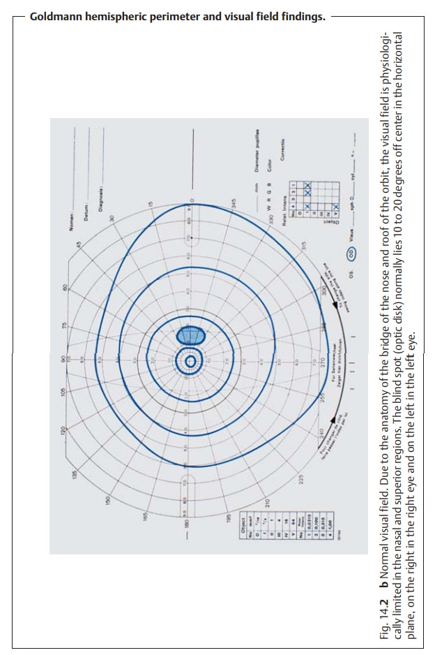
The points of light decrease in size and light intensity as they move toward
the center of the visual field, and the isopters become correspond-ingly
smaller (Fig. 14.2b). This
corresponds with the sensitivity of the retina, which increases from the
periphery to the center.
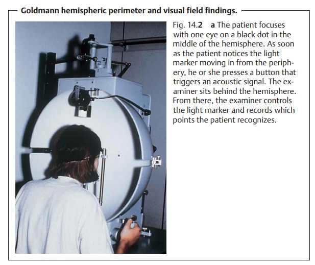
The advantage of kinetic perimetry is the
personal interaction between physician and patient. This method is especially
suitable for older patients who may have difficulties with a stereotyped
interaction required by a com-puter program. Specific indications for kinetic perimetry include visual field defects
due to neurologic causes and examinations to establish a disability (such as
hemianopsia or quadrantic anopsia).
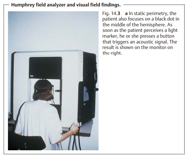
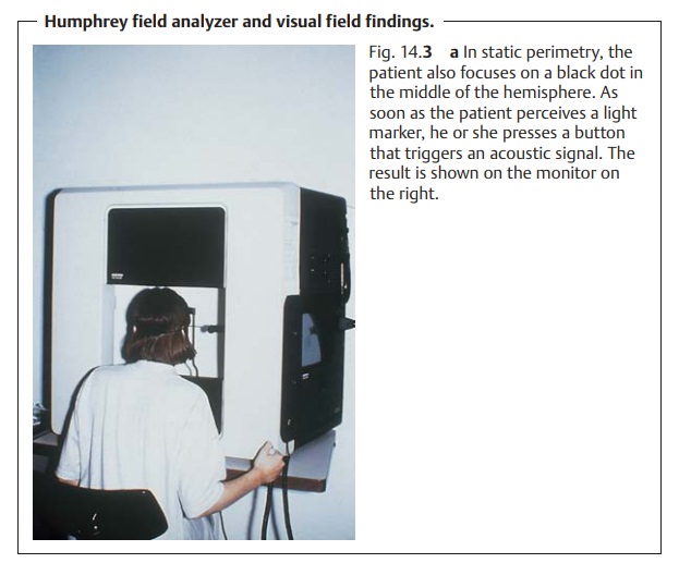
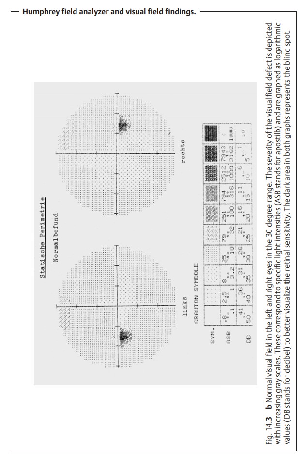
2. Static perimetry.
This is usually performed with computerized equipmentsuch as the
Humphrey field analyzer (Fig. 14.3) or Octopus 2000, although a Goldmann or Rodenstock hemispheric
perimeter can also be used for static testing of the visual field. In static
perimetry, the light intensity of immobile
light markers is increased until they are perceived. The intensity threshold
continuously increases from the macula, with the highest sensitivity, to the
periphery. A variety of different computer programs can be selected depend-ing
on the specific clinical setting. These include the outer margins or the 30
degree visual field in glaucoma (Fig. 14.3b).
Other examination methods:
❖Pupillary findings.
❖Pupillary light reflex.
❖ Visual
evoked potential.
❖ CT or MRI to diagnose causes.
Related Topics