Chapter: The Massage Connection ANATOMY AND PHYSIOLOGY : Urinary System
Urination or Micturition
Urination or Micturition
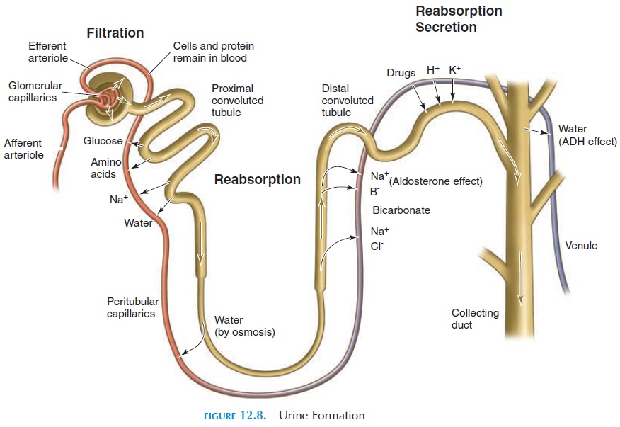
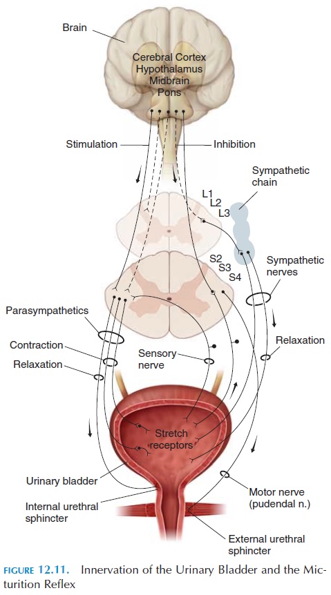
As the bladder fills with urine, the walls are stretched and the stretch receptors here are stimulated. Impulses are conducted by sensory nerves (pelvic nerves) to the sacral segment (S2 and S3), the micturition center (Figure 12.8).

The nerves synapse here with parasympathetic nerves, whose action is to contract the detrusor and relax the internal sphincter. The micturition center also communicates with the motor nerves that inner-vate the skeletal muscles of the external urethral sphincter. The sensory nerves communicate with other nerves that carry impulses to the thalamus, the brainstem, and other areas of the cerebral cortex. The latter is responsible for the conscious awareness of a filled bladder. Communication from higher centers to the micturition center facilitates or inhibits the mic-turition reflex. Usually, the urge to urinate begins when the bladder is filled with about 200 mL (12.2 in3) of urine (see Figure 12.11.)

Both the internal (involuntary control) and exter-nal urethral sphincters (voluntary control) must relax for the bladder to be emptied. Infants lack voluntary control, and the bladder is emptied reflexively when the bladder becomes distended. Therefore, micturi-tion is a spinal reflex that can be facilitated or inhib-ited by higher brain centers. The ability to keep the external urethral sphincter contracted and delay uri-nation is a learned process.
After urination, the female urethra empties by gravity; in males, it empties by contraction of the bulbocavernosus muscle.
Related Topics