Chapter: The Massage Connection ANATOMY AND PHYSIOLOGY : Urinary System
The Kidneys
THE KIDNEYS
The paired kidneys (Figure 12.2) are bean-shaped, with the indentation (the hilum) facing medially. The renal artery, renal vein, lymphatics, and renal nerves
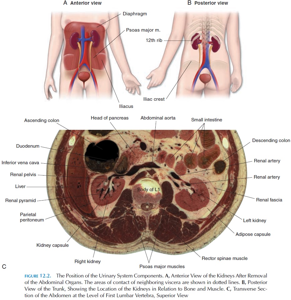
The kidneys are located on either side of the vertebral column between the T12 and L3 vertebrae. The right kidney is slightly lower than the left because of the presence of the liver. These organs are retroperitoneal (i.e., they are located behind the peritoneum). The anterior aspect of the right kidney is related to the liver, the hepatic flexureof the colon, and the duodenum. The left kidney is covered by the stomach, pancreas, spleen, jejunum, and splenic flexure of the colon. The superior aspect of both the kidneys is covered by the adrenal glands.
Posteriorly, the kidneys are related to the muscles of the back; the tendon of the transverses abdominis muscle, the quadratus lumborum and the psoas, the diaphragm, the eleventh and twelfth ribs on the left side and the twelfth rib on the right.
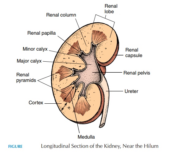
The kidneys are held in place, supported, and pro-tected by the surrounding fat and connective tissue. From deep to superficial, the kidney is invested by a fibrous layer of dense collagen fibers known as the re-nal capsule. The renal capsule forms a smooth andfirm covering to the organ. Medially, the renal cap-sule folds inward at the hilus and lines an internal cavity, the renal sinus. The renal blood vessels and

The kidney and its ves-sels are embedded in a mass of adipose tissue (adi-pose capsule, or perirenal fat). Both the kidneys and the fatty tissue are surrounded by a thick outer layer of fascia, the renal fascia, which anchors the kidneys to surrounding structures such as the peri-toneum anteriorly and the fascia covering the back muscles posteriorly. Because the kidneys are not di-rectly fixed to the abdominal wall, they move with the diaphragm during respiration.
If a section is made through the kidney, it is found to have two distinctive parts—the outer cortex and an inner medulla. Medially, the medulla forms 6–18 conical structures known as renal pyramids. The part of the cortex that dips between the pyramids is the renal column. The apex of the pyramids—the re-nal papilla, located medially—projects into the renalsinus. Urine that is formed is drained by the ducts in the papilla into cup-shaped structures known as re-nal calyces (singular, calyx). Four or five smaller ca-lyces—theminor calyces—empty into two to three larger, major calyces. The major calyces join and form a large chamber, the renal pelvis.The funnel-shaped pelvis, which occupies most of the renal si-nus, is continuous with the ureter.
Related Topics