Chapter: Modern Pharmacology with Clinical Applications: General Organization and Functions of the Nervous System
Transmission of the Nerve Impulse

TRANSMISSION OF
THE NERVE IMPULSE
Microscopic studies of the
structure of the terminal axons of the autonomic nerves have shown that the
ax-ons branch many times on entering the effector tissue, forming a plexus
among the innervated cells. “Swollen” areas found at intervals along the
terminal axons are re-ferred to as varicosities
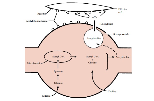
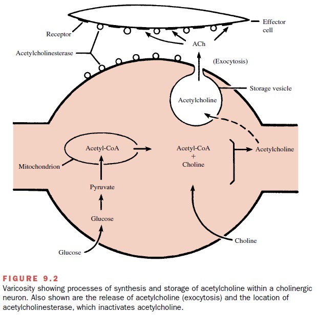
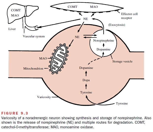
(Figs. 9.2 and 9.3). Within each varicosity are mitochondria and numerous vesicles con-taining neurotransmitters.


The vesicles are intimately
involved in the release of the transmitter into the synaptic or neuroeffector
cleft in response to an action potential. Following release, the
transmitter must diffuse to the effector cells, where it in-teracts with
receptors on these cells to produce a re-sponse. The distance between the
varicosities and the effector cells varies considerably from tissue to tissue.
Smooth muscle, cardiac muscle, and exocrine gland cells do not contain
morphologically specialized regions comparable to the end plate of skeletal
muscle.
In the autonomic ganglia, the
varicosities in the terminal branches of the preganglionic axons come into
close contact primarily with the dendrites of the ganglionic cells and make
synaptic connection with them.
Related Topics