Chapter: Maternal and Child Health Nursing : Urinary System
The ureters
The ureters
These are
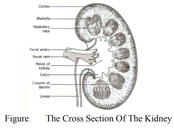
the tubes that convey urine from the kidneys to the urinary bladder; the upper
end is funnel-shape where it merges with the pelvis of the kidneys. Each tube
is about 25-30cm long 1 and 3mm in diameter. It runs from the renal hilum to
the posterior
wall of
the bladder, pass down the posterior wall of the abdomen towards the pelvis, so
remain outside the peritoneal cavity, beside the uterine cervix to enter the
bladder from behind.
Structure
The ureter
is made up of three layers.
·
Transitional
Epithelium: This forms the lining of theureter and is arranged
in longitudinal folds.
·
Muscular
Layer: This layer is composed of three fibers;inner longitudinal fibers, middle
circular fibers and outer longitudinal fibers.
·
Outer
Coat: This is made up of fibrous connective tissue which is continuous with
the fibrous tissue of the kidneys. It is a protective layer.
Blood Supply
The upper
part: by the renal artery in the pelvic portion- Common Iliac, internal iliac,
uterine and vesical arteries, Venous Returns- Corresponding veins.
Lymphatic
Drainage- Internal and external common iliac lymph nodes.
Nerve
Supply- Aortic, Renal superior and inferior hyponastic plexus. Sympathetic and
parasympathetic nerves.
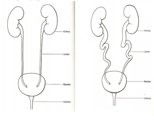
Effect of Pregnancy on the Ureters
Pregnancy
hormone e.g. progesterone relaxes the walls of the ureters. This results in
dilatation and linking of the ureters which tends to slow down the flow of
urine or causes stasis. This increases the risk of infection in pregnancy.
Related Topics