Chapter: Human Neuroanatomy(Fundamental and Clinical): The Olfactory and Limbic Regions
The Olfactory Pathway
The Olfactory Pathway
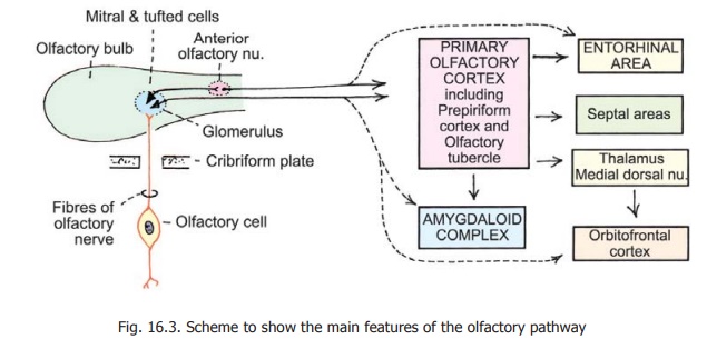
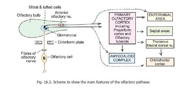
The fibres of the olfactory nerves are processes of olfactory receptor cells lying in the epithelium lining the olfactory mucosa (Fig. 16.3). These receptor cells are homologous to sensory neurons located in sensory ganglia. In other words, the first order sensory neurons of the olfactory pathway are located within the olfactory epithelium itself. (In the course of evolution all neurons have arisen by modification of epithelial cells and their migration, in most cases, into deeper tissues. The olfactory receptor cells retain their position in the epithelium and are, therefore, regarded as primitive).
Each receptor cell consists of a cell body and of two processes i.e., it is a bipolar cell. The peripheral process (dendrite) reaches the surface of the olfactory epithelium and ends in a small swelling. A number of cilia are attached to this swelling. The central process (axon) enters the submucosa, and forms one fibre of the olfactory nerve. The olfactory nerve fibres terminate in the olfactory bulb.
Olfactory Bulb
The olfactory bulb receives fibres of the olfactory nerves, arising from olfactory cells (or olfactory sensory neurons). These incoming fibres synapse with neurons within the bulb. Fibres arising from the latter form the olfactory tract.
Several types of cells are present in the olfactory bulb. The most important of these are:
a. The mitral cells and tufted cells that give origin to fibres of the olfactory tract.
Advanced:
Other cells present are as follows:
b. Periglomerular cells and granule cells have processes that remain confined to the bulb.
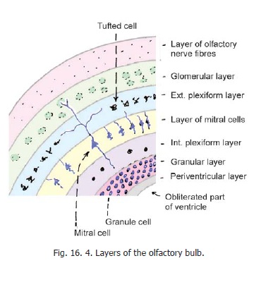
The olfactory bulb is made up of a number of concentric layers which are shown in Fig. 16.4.
1. Incoming fibres of olfactory nerves occupy the most superficial layer.
2. The second, or glomerular, layer contains synaptic glomeruli in which terminals of olfactory nerve fibres synapse with dendrites of mitral cells, tufted cells and periglomerular cells.
3. The third layer is in the form of a plexus formed by dendrites of mitral and tufted cells. Somata of tufted cells also lie in this layer.
4. The fourth layer contains somata of mitral cells.
5. The fifth layer is made up mainly of axons of mitral and tufted cells.
6. The sixth layer contains clusters of granule cells.
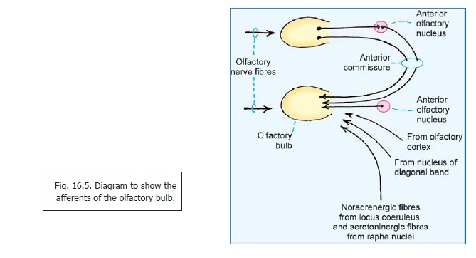
Apart from olfactory nerve fibres, the olfactory bulb receives centrifugal fibres from a number of sources as shown in Fig. 16.5. It is believed that perception of smell can be considerably modulated through these inputs.
Many neuropeptides are present in the olfactory bulb. These include GABA, dopamine, glutamate, aspartate, enkephalin, luteinising hormone releasing hormone (LHRH) and substance P.
Olfactory tract
The olfactory tract is made up predominantly of axons of mitral and tufted cells of the olfactory bulb. Some scattered neurons are also present within the tract. They collectively constitute theanterior olfactory nucleus. Some axons of mitral and tufted cells relay in this nucleus.
The olfactory tract also contains centrifugal fibres travelling to the olfactory bulb from various centres in the brain. These are shown in Fig. 16.5.
Olfactory cortex
The term olfactory cortex is applied to all areas of the cerebral cortex that receive direct fibres from the olfactory bulb.
In the past it has been held that olfactory pathways differ from those for other sensations in that there is no relay in the thalamus. This concept needs modification in that while most olfactory fibres reach their cortical destinations without relay (ignoring relay of some in the anterior olfactory nucleus), some others are now believed to reach the thalamus.
Primary Olfactory Projection
The main regions receiving direct fibres from the olfactory bulb are:
1. The prepiriform cortex (including the lateral olfactory gyrus and the gyrus ambiens).
2. The gyrus semilunaris (periamygdaloid area). Some direct fibres also reach the following:
3. Some direct fibres also reach the following:
4. Olfactory tubercle (in the anterior perforated substance).
5. Entorhinal area.
The term piriform cortex or primary olfactory cortex is rather loosely applied to these regions, the areas included varying in different accounts. Apart from the above, some direct olfactory axons also reach some parts of the cortex of the insula; and the main (central) nuclei of the amygdaloid complex.
Secondary olfactory projection
1. Apart from primary olfactory fibres the entorhinal area receives numerous olfactory fibres afterrelay in other parts of the piriform cortex.
2. It has been recently recognised that the piriform cortex sends a major projection to the medialdorsal nucleus of the thalamus. From here olfactory impulses are relayed to the cortex on the orbital aspect of the frontal lobe (orbito-frontal cortex, centro-posterior part)
3. Another region of the orbitofrontal cortex (latero-posterior) receives direct projections from thepiriform cortex. This projection by-passes the thalamus. Studies on alterations in brain blood flow in response to olfactory stimuli suggest that appreciation of such stimuli takes place bilaterally in the piriform cortex, but only in the right frontal lobe.
The primary olfactory cortex is connected to various other areas. In the past, many of these areas have been included under the term rhinencephalon or smell brain. It is now recognised, however, that although these areas might receive olfactory impulses their primary functions are not olfactory. Some of these areas and their connections are considered below as part of the limbic system.
Related Topics