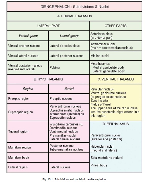
Chapter: Human Neuroanatomy(Fundamental and Clinical): The Olfactory and Limbic Regions
Fibre bundles of limbic region
Fibre bundles of limbic region
Stria Terminalis
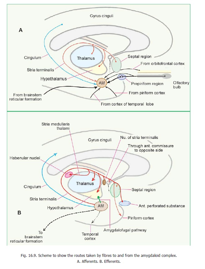
This bundle of fibres is closely related to the inferior horn and central part of the lateral ventricle. It begins in the amygdaloid complex and runs backwards in the roof of the inferior horn. It then winds upwards and forwards to lie in the floor of the central part of the ventricle. Finally, it terminates near the interventricular foramen and anterior commissure by dividing into various smaller bundles. Throughout its course, it is closely related to the medial side of the caudate nucleus (Fig. 13.1). In the inferior horn it is related to the tail of this nucleus. In the central part of the ventricle it lies medial to the body of the caudate nucleus. Here the thalamus is medial to it. A group of neurons located just behind the anterior commissure is sometimes described as the nucleus of the stria terminalis (Fig. 16.9B).
Advanced:
The stria terminalis contains the following fibres (as traditionally described, Figs. 16.9 and 16.11).
1. Fibres from the amygdaloid complex to the septal region, the hypothalamus, the anteriorperforated substance, the piriform lobe and the nucleus of the stria terminalis.
2. Fibres from the amygdaloid nucleus to the habenular nuclei. These fibres leave the stria at itsanterior end and reach the habenular nuclei through the stria medullaris thalami.
3. Some fibres from the amygdaloid complex run forwards in the stria, cross to the oppositeside in the anterior commissure, and then run backwards in the stria of the opposite side to reach the opposite amygdaloid complex.
4. Fibres from the septal areas and adjoining regions possibly run backwards in the stria to reachthe amygdaloid complex.
Anterior Commissure
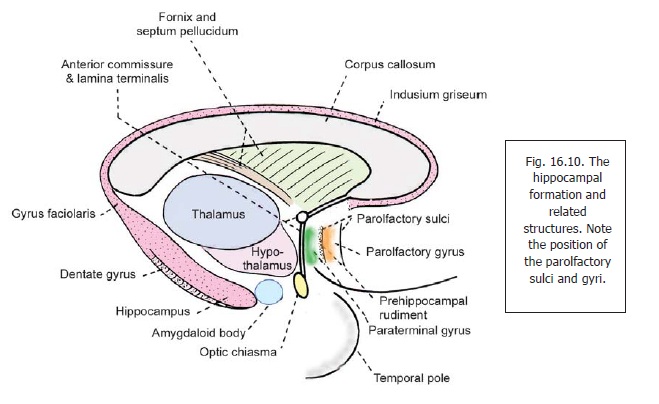
The anterior commissure is situated in the anterior wall of the third ventricle at the upper end of the lamina terminalis (Fig. 16.10).
Advanced:
When traced laterally, it divides into anterior and posterior bundles. The posterior bundle passes below the lentiform nucleus. Fibres passing through the commissure interconnect the regions of the two cerebral hemispheres concerned with the olfactory pathway. These include the olfactory bulb, the anterior olfactory nucleus, the prepiriform cortex, the entorhinal area, the anterior perforated substance, the amygdaloid complex and related structures. Other fibres interconnect the parahippocampal gyri and other parts of the two temporal lobes. Some fibres interconnect the frontal lobes of the two sides.
The Fornix
The fornix is a prominent bundle of fibres seen on the medial aspect of the cerebral hemisphere. It is made up, predominantly, of fibres arising in the hippocampus. The body of the fornix is suspended from the corpus callosum by the septum pellucidum (Figs. 13.1, 8.6) and comes into close relationship with the tela choroidea in the roof of the third ventricle. When traced posteriorly, the body of the fornix divides into two parts called crura. Each crus of the fornix becomes continuous with the fimbria of the corresponding side. The two crura are interconnected by fibres passing from one to the other. These crossing fibres constitute the hippocampal commissure or commissure of thefornix. The anterior end of the body of the fornix also divides into right and left halves called the columns of the fornix. Each column turns downwards just in front of the interventricular foramenand passes through the hypothalamus to reach the mamillary body (Fig. 16.15B).
Advanced:
Most of the fibres of the fornix lie behind the anterior commissure and are, therefore, referred to as the postcommissural fornix. In contrast some fibres of the fornix that descend in front of the anterior commissure constitute the precommissural fornix. Some of the fibres of the fornix pass above the splenium of the corpus callosum to reach structures above the latter. These fibres constitute the dorsal fornix.
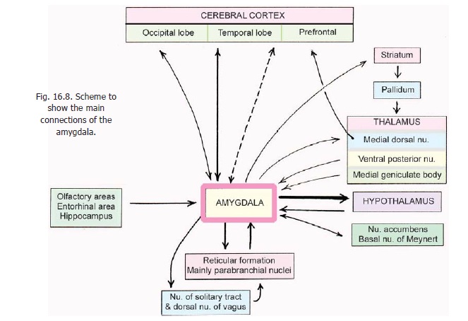
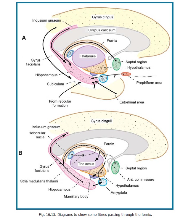
The fibres contained in the fornix are as follows (Fig. 16.8):
1. Fibres interconnecting the hippocampi of the two sides through the hippocampal commissure.
2. Fibres from the hippocampus that pass through the postcommissural fornix to reach the mamillarybody. These are then relayed to the anterior nucleus of the thalamus. Some fibres of the fornix end directly in this nucleus and some in the hypothalamus.
3. Fibres from the hippocampus that pass through the precommissural fornix reach the septalregion. Some of these fibres turn backwards to enter the stria medullaris thalami and reach the habenular nuclei.
4. Fibres entering the dorsal fornix reach the splenial gyrus and the gyrus cinguli.
Related Topics