The Cell | Term 2 Unit 5 | 6th Science - Student Activities | 6th Science : Term 2 Unit 5 : The Cell
Chapter: 6th Science : Term 2 Unit 5 : The Cell
Student Activities
Activity 1:
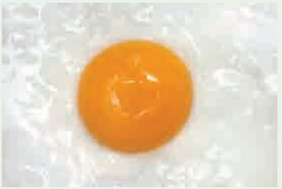
Aim: To observe the structure of a single cell (Hen’s egg).
Materials Needed: A hen’s egg and a plate.
Method: Crack the shell and break open the egg in a plate.
Observation: The egg has a yellow part and a transparent part surrounding it. The white transparent part (albumin) is jelly-like and represents the cell’s cytoplasm, while the yellow part (yolk) is thicker and represents the cell’s nucleus. On the internal side of the shell can be seen a thin membrane-like structure, which represents the cell membrane.

Activity 2:
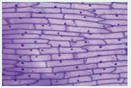
Aim: To observe onion peel cells under a microscope
Materials Required: Glass slide, cover slip, onion, iodine solution, knife and microscope.
Procedure: Take an onion and cut it into two halves along its length. Take out one of its fleshy leaves. With the help of a pair of forceps, remove a transparent, thin peel from the inner surface of the leaf. Take a glass slide and put a drop of water at the centre. Place the peel on the drop of water. Pour a drop of iodine solution on the peel. Now place a cover slip over the material. Observe under the microscope.
Observation: You will be able to see rectangular cells of the onion peel, with a nucleus in each of them.

Activity 3:
Aim:
To rectify the variation between 2-D shape and 3-D shape.
Material required:
Polythene bag, water, marble ball (golli gundu)
Procedure:
Take a polythene bag with water. Put a marble ball into the polythene bag. Then draw a picture in your note book about this task.
If you draw a picture in round shape.
It will be called 2-Dimenstional picture.
If you draw a picture in spherical shape it is called 3-dimenstional.
Result:
Now you understand your misconceptions. So the animal cells are spherical in shape and structure, not in a round shape.
Related Topics