Chapter: Human Neuroanatomy(Fundamental and Clinical): Tracts of Spinal Cord and Brainstem
Spinocerebellar Pathways - Tracts of Spinal Cord and Brainstem
Spinocerebellar Pathways
These pathways carry proprioceptive impulses arising in muscle spindles, Golgi tendon organs, and other receptors to the cerebellum. They constitute the afferent component of reflex arcs involving the cerebellum, for control of posture. Recent investigations have shown that some exteroceptive sensations (e.g., touch) may reach the cerebellum through these pathways.
1. The first order neurons of these pathways are located in dorsal nerve root ganglia. Their peripheral processes end in relation to muscle spindles, Golgi tendon organs and other proprioceptive receptors. Some fibres are related to end organs concerned with exteroceptive sensations (touch, pressure). The central processes of the neurons concerned ascend in the posterior funiculi for varying distances before ending in spinal grey matter. Some of them ascend all the way to the medulla and end in the accessory cuneate nucleus.
2. The second order neurons of the pathway are arranged in a number of groups.
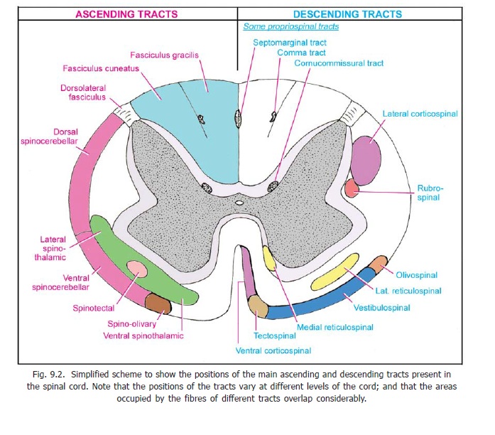
i. Neurons located in the dorsal nucleus (situated on the medial side of the base of the dorsal grey column in segments C8 to L3 of the spinal cord: Fig. 5.2) give origin to fibres of the dorsal (posterior)spinocerebellar tract. This is an uncrossed tract lying in the lateral funiculus (Fig. 9.2). It begins inthe lumbar segments of the spinal cord and ascends to the medulla where its fibres become incorporated in the inferior cerebellar peduncle and pass through it to reach the vermis of the cerebellum (Fig. 9.8).
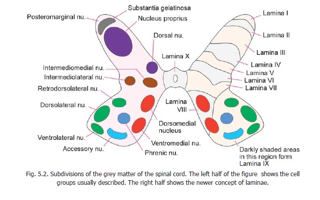
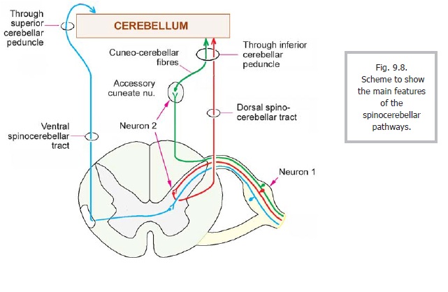
ii. The neurons giving origin to the ventral (anterior) spinocerebellar tract are probably located in the junctional area between the ventral and dorsal grey columns (laminae V, VI, VII) in the lumbar and sacral segments of the cord. Some of the neurons concerned may lie in the ventral grey column (spinal border cells). The fibres of this tract are predominantly crossed. They ascend in the lateral funiculus, anterior to the fibres of the dorsal spinocerebellar tract (Fig. 9.2), and pass through the medulla and pons. At the upper end of the pons the fibres turn downwards to enter the superior cerebellar peduncle through which they reach the vermis of the cerebellum (Fig. 9.8).From a functional point of view both the ventral and dorsal spinocerebellar tracts are concerned mainly with the lower limbs and trunk. The dorsal tract is believed to carry impulses concerned with fine co-ordination of muscles controlling posture, and with movements of individual muscles. On the other hand the ventral tract is concerned with movements of the limb as a whole.
iii. We have seen that the central processes of some first order neurons (related to cervical segments) reach the accessory cuneate nucleus (also called the lateral or external cuneate nucleus) in the medulla. Second order neurons lying in this nucleus give origin to posterior external arcuatefibres which enter the inferior cerebellar peduncle (of the same side) to reach the cerebellum. Thiscuneocerebellar tract carries impulses from the upper limb. It may be regarded as the forelimbequivalent of the dorsal spinocerebellar tract.
iv. Rostral spinocerebellar pathway: This tract is believed to arise from spinal grey matter in cervical regions of the spinal cord. The neurons of origin lie in the lower four cervical segments (lamina VII). These neurons constitute the nucleus centrobasalis. Most fibres of the pathway are uncrossed. They reach the cerebellum through the inferior and superior cerebellar peduncles. This pathway is regarded, functionally, as the forelimb equivalent of the ventral spinocerebellar tract. From the above account of spinocerebellar pathways it will be seen that these are two neuron pathways in contrast to three neuron pathways to the cerebral cortex.
v. Impulses from the spinal cord may reach the cerebellum indirectly by traveling through the spino-olivary tract, and through olivocerebellar fibres. Similarly, impulses may reach the cerebellum after traveling through spinoreticular and reticulocerebellar fibres.
vi. A cervicocerebellar pathway carrying proprioceptive impulses from neck muscles has been described in some animals. The neurons of origin lie in the same region as those of the rostral cerebellar pathway, but in the upper four cervical segments. The fibres pass through the superior cerebellar peduncle to reach the cerebellum.
Spinocerebellar fibres reaching the cerebellum end as mossy fibres. Representation in the cerebellar cortex is somatotropic (i.e., fibres carrying sensations from different parts of the body end in definite areas of cortex). Conduction in the spinocerebellar tracts can be inhibited or facilitated under the influence of descending tracts (corticospinal, reticulospinal, rubrospinal).
Related Topics