Chapter: Medicine and surgery: Cardiovascular system
Signs of Jugular venous pressure
Jugular venous pressure
The internal jugular vein is most easily seen with the patient reclining
(usually at 45Ëš), with the head supported and the neck muscles relaxed and in
good lighting conditions. The jugular vein runs medial to the sternomastoid
muscle in the upper third of the neck, behind it in the middle third and
between the two heads of sternocleidomastoid in the lower third. It is
differentiated from the carotid pulse by its double waveform, it is
non-palpable, it is occluded by pressure and pressure on the liver causes a
rise in the level of the pulsation (hepato-jugular reflex). The jugular
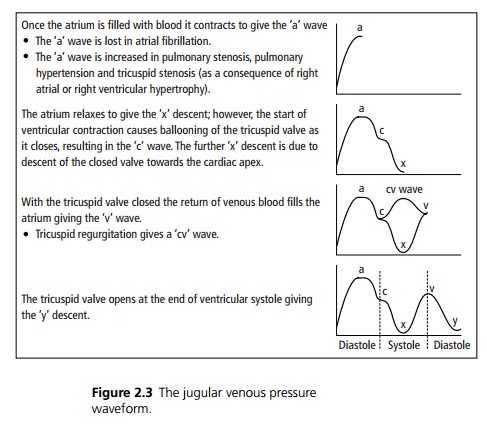
waveform and pressure give information about the pressures within the right
atrium as there are no valves separating the atrium and the internal jugular
vein (see Fig. 2.3).
The height of the jugular venous pressure (JVP) is assessed as the
vertical height from the sternal angle to the point at which the JVP is seen. A
height of greater than 3 cm represents an abnormal increase in filling pressure
of the right atrium. This may occur in right-sided heart failure, congestive
cardiac failure and pulmonary embolism.
Related Topics