Chapter: Essentials of Anatomy and Physiology: The Heart
Regulation of Heart Rate
REGULATION OF HEART RATE
Although the heart generates and maintains its own beat, the rate of contraction can be changed to adapt to different situations. The nervous system can and does bring about necessary changes in heart rate as well as in force of contraction.
The medulla of the brain contains the two cardiac centers, the accelerator center and the inhibitory center. These centers send impulses to the heart along autonomic nerves. Recall that the autonomic nervous system has two divisions: sym-pathetic and parasympathetic. Sympathetic impulses from the accelerator center along sympathetic nerves increase heart rate and force of contraction during exercise and stressful situations. Parasympathetic impulses from the inhibitory center along the vagus nerves decrease the heart rate. At rest these impulses slow down the depolarization of the SA node to what we consider a normal resting rate, and they also slow the heart after exercise is over.
Our next question might be: What information is received by the medulla to initiate changes? Because the heart pumps blood, it is essential to maintain nor-mal blood pressure. Blood contains oxygen, which all tissues must receive continuously. Therefore, changes in blood pressure and oxygen level of the blood are stimuli for changes in heart rate.
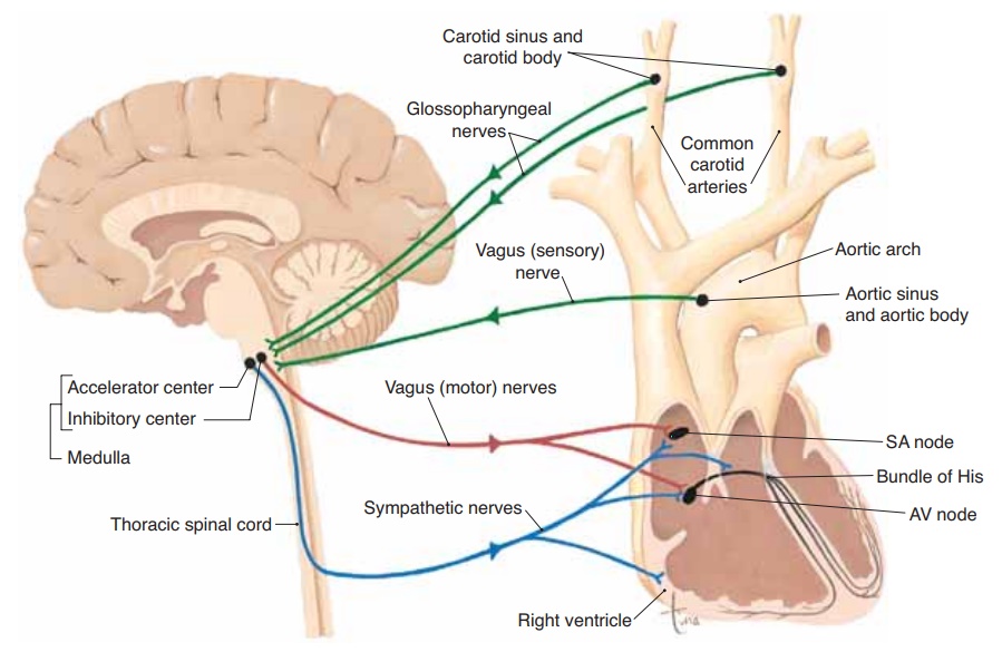
You may also recall that presso-receptors and chemoreceptors are located in the carotid arteries and aortic arch. Pressoreceptors in the carotid sinuses and aortic sinus detect changes in blood pressure. Chemoreceptors in the carotid bodies and aortic body detect changes in the oxygen content of the blood. The sensory nerves for the carotid receptors are the glossopharyngeal (9th cra-nial) nerves; the sensory nerves for the aortic arch receptors are the vagus (10th cranial) nerves. If we now put all of these facts together in a specific exam-ple, you will see that the regulation of heart rate is a reflex. Figure 12–7 depicts all of the structures just mentioned.
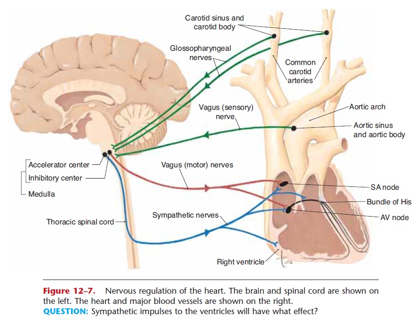
Figure 12–7. Nervous regulation of the heart. The brain and spinal cord are shown on the left. The heart and major blood vessels are shown on the right.
QUESTION: Sympathetic impulses to the ventricles will have what effect?
A person who stands up suddenly from a lying posi-tion may feel light-headed or dizzy for a few moments, because blood pressure to the brain has decreased abruptly. The drop in blood pressure is detected by pressoreceptors in the carotid sinuses—notice that they are “on the way” to the brain, a very strategic location. The drop in blood pressure causes fewer impulses to be generated by the pressoreceptors. These impulses travel along the glossopharyngeal nerves to the medulla, and the decrease in the fre-quency of impulses stimulates the accelerator center. The accelerator center generates impulses that are car-ried by sympathetic nerves to the SA node, AV node, and ventricular myocardium. As heart rate and force increase, blood pressure to the brain is raised to nor-mal, and the sensation of light-headedness passes.
When blood pressure to the brain is restored to nor-mal, the heart receives more parasympathetic impulses from the inhibitory center along the vagus nerves to the SA node and AV node. These parasympathetic impulses slow the heart rate to a normal resting pace.
The heart will also be the effector in a reflex stim-ulated by a decrease in the oxygen content of the blood. The aortic receptors are strategically located so as to detect such an important change as soon as blood leaves the heart. The reflex arc in this situation would be (1) aortic chemoreceptors, (2) vagus nerves (sen-sory), (3) accelerator center in the medulla, (4) sympa-thetic nerves, and (5) the heart muscle, which will increase its rate and force of contraction to circulate more oxygen to correct the hypoxia.
Recall that the hormone epinephrine is secreted by the adrenal medulla in stressful situations. One of the many functions of epi-nephrine is to increase heart rate and force of contrac-tion. This will help supply more blood to tissues in need of more oxygen.
Related Topics