Chapter: Essentials of Anatomy and Physiology: The Heart
Cardiac Conduction Pathway
CARDIAC CONDUCTION PATHWAY
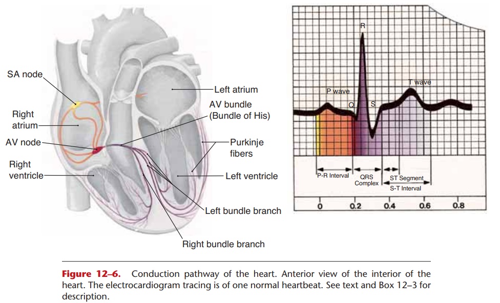
The cardiac cycle is a sequence of mechanical events that is regulated by the electrical activity of the myocardium. Cardiac muscle cells have the ability to contract spontaneously; that is, nerve impulses are not required to cause contraction. The heart generates its own beat, and the electrical impulses follow a very specific route throughout the myocardium. You may find it helpful to refer to Fig. 12–6 as you read the following.
The natural pacemaker of the heart is the sinoatrial (SA) node, a specialized group of cardiac muscle cells located in the wall of the right atrium just below the opening of the superior vena cava. The SA node is considered specialized because it has the most rapid rate of contraction, that is, it depolarizes more rapidly than any other part of the myocardium (60 to 80 times per minute). As you may recall, depolarization is the rapid entry of Na+ ions and the reversal of charges on either side of the cell membrane. The cells of the SA node are more permeable to Na+ ions than are other cardiac muscle cells. Therefore, they depolarize more rapidly, then contract and initiate each heartbeat.
From the SA node, impulses for contraction travel to the atrioventricular (AV) node, located in the lower interatrial septum. The transmission of impulses from the SA node to the AV node and to the rest of the atrial myocardium brings about atrial systole.
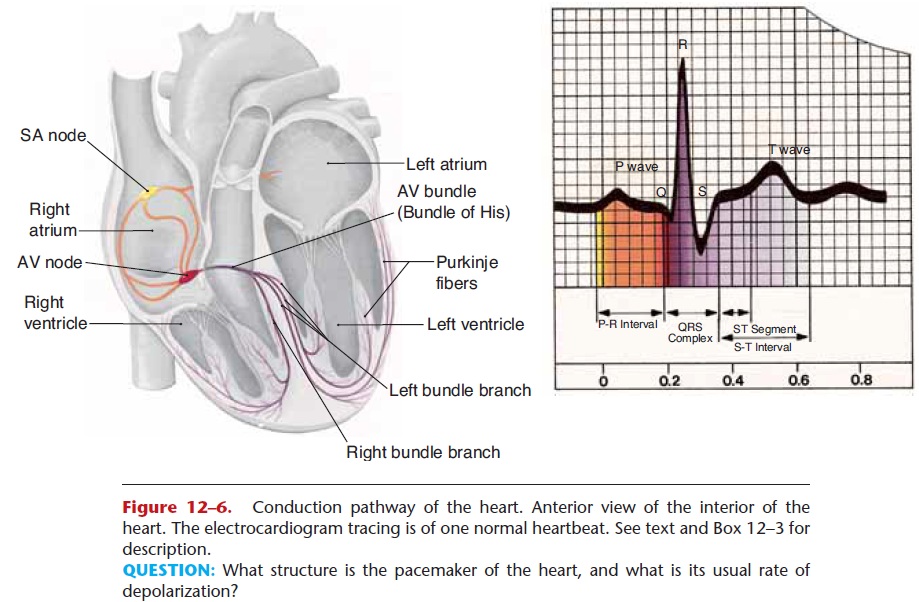
Figure 12–6. Conduction pathway of the heart. Anterior view of the interior of the heart. The electrocardiogram tracing is of one normal heartbeat.
QUESTION: What structure is the pacemaker of the heart, and what is its usual rate of depolarization?
Recall that the fibrous skeleton of the heart sepa-rates the atrial myocardium from the ventricular myocardium; the fibrous connective tissue acts as elec-trical insulation between the two sets of chambers. The only pathway for impulses from the atria to the ventricles, therefore, is the atrioventricular bundle (AV bundle), also called the bundle of His. The AV bundle is within the upper interventricular septum; it receives impulses from the AV node and transmits them to the right and left bundle branches. From the bundle branches, impulses travel along Purkinje fibers to the rest of the ventricular myocardium and bring about ventricular systole. The electrical activity of the atria and ventricles is depicted by an electrocar-diogram (ECG); this is discussed in
If the SA node does not function properly, the AV node will initiate the heartbeat, but at a slower rate (50 to 60 beats per minute). The AV bundle is also capa-ble of generating the beat of the ventricles, but at a much slower rate (15 to 40 beats per minute). This may occur in certain kinds of heart disease in which transmission of impulses from the atria to the ventri-cles is blocked.
Arrhythmias are irregular heartbeats; their effects range from harmless to life-threatening. Nearly everyone experiences heartpalpitations (becoming aware of an irregular beat) from time to time. These are usually not serious and may be the result of too much caffeine, nicotine, or alcohol. Much more serious is ventricular fibrillation, a very rapid and uncoordinated ventricular beat that is totally inef fective for pumping blood.
Related Topics