Principle, Working Mechanism, Applications, Optical Components - Phase Contrast Microscope | 12th Microbiology : Chapter 2 : Microscopy
Chapter: 12th Microbiology : Chapter 2 : Microscopy
Phase Contrast Microscope
Phase Contrast Microscope.
Frits
Zernike a Dutch Physicist invented the Phase Contrast Microscope and was
awarded Nobel Prize in 1953. It is the microscope which allows the observation
of living cell. This microscopy uses special optical components to exploit fine
differences in the refractive indices of water and cytoplasmic components of
living cells to produce contrast.
Principle
The phase
contrast microscopy is based on the principle that small phase changes in the
light rays, induced by differences in the thickness and refractive index of the
different parts of an object, can be transformed into differences in brightness
or light intensity. The phase changes are not detectable to human eye whereas the
brightness or light intensity can be easily detected.
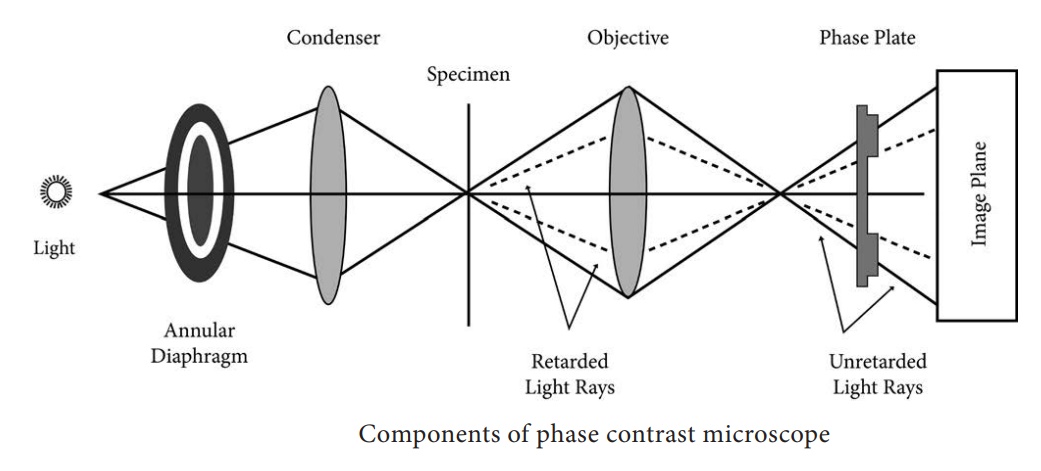
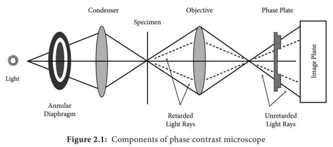
Optical Components of Phase Contrast Microscope (PCM)
The phase
contrast microscope is similar to an ordinary compound microscope in its
optical components. It possesses a light source, condenser system, objective
lens system and ocular lens system (Figure 2.1).
A phase
contrast microscope differs from bright field microscope in having,
i. Sub-stage annular diaphragm (phase condenser)
An
annular aperture in the diaphragm is placed in the focal plane of the sub-stage
which controls the illumination of the object. This is located below the
condenser of the microscope. This annular diaphragm helps to create a narrow,
hollow cone of light to illuminate the object.
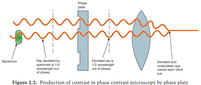
ii. Phase – plate (diffraction plate or phase retardation plate)
This
plate is located at the back focal plane of the objective lenses. The phase
plate has two portions, in which one is coated with light retarding material
(Magnesium fluoride) and the other portion devoid of light retarding material
but can absorb light. This plate helps to reduce the phase of the incident
light (Figure 2.2).
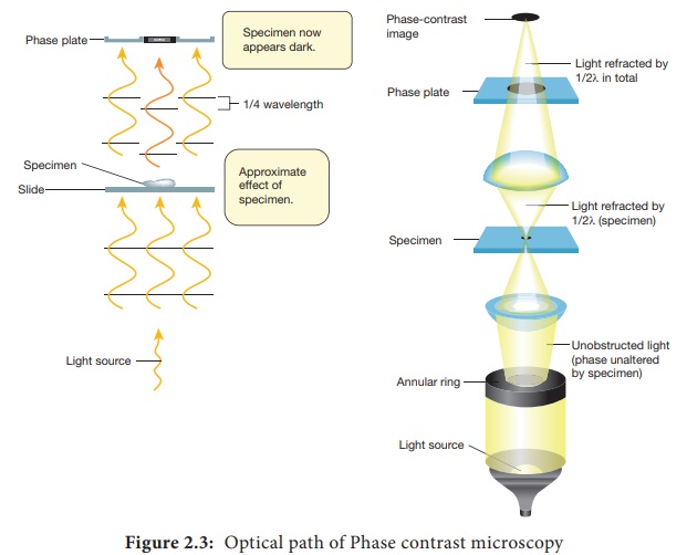
Working Mechanism of Phase Contrast Microscopy
The
unstained cells cannot create contrast under the normal microscope. However,
when the light passes through an unstained cell, it encounters regions in the
cell with different refractive indexes and thickness. When light rays pass
through an area of high refractive index, it deviates from its normal path and
such light rays experience phase change or phase retardation (deviation). Light
rays pass through the area of less refractive index remain non-deviated (no
phase change). Figure 2.3 shows the light path in phase contrast microscope
HOTS: How does phase contrast
microscope differ from Bright Field microscope?
The
difference in the phases between the retarded (deviated) and un-retarded
(non-deviated) light rays is about ¼ of original wave length (i.e., λ/4). Human
eyes cannot detect these minute changes in the phase of light. The phase
contrast microscope has special devices such as annular diaphragm and phase
plate, which convert these minute phase changes into brightness (amplitude)
changes, so that a contrast difference can be created in the final image. This
contrast difference can be easily detected by human eyes.
In phase
contrast microscope, to get contrast, the diffracted waves have to be separated
from the direct waves. This separation is achieved by the sub-stage annular
diaphragm.
The
annular diaphragm illuminates the specimen with a hollow cone of light. Some
rays (direct rays) pass through the thinner region of the specimen and do not
undergo any deviation and they directly enter into the objective lens. The
light rays passing through the denser region of the specimen get regarded and
they run with a delayed phase than the non-deviated rays. Both the deviated and
non deviated light has to pass through the phase plate kept on the back focal
plane of the objective to form the final image. The difference in phase
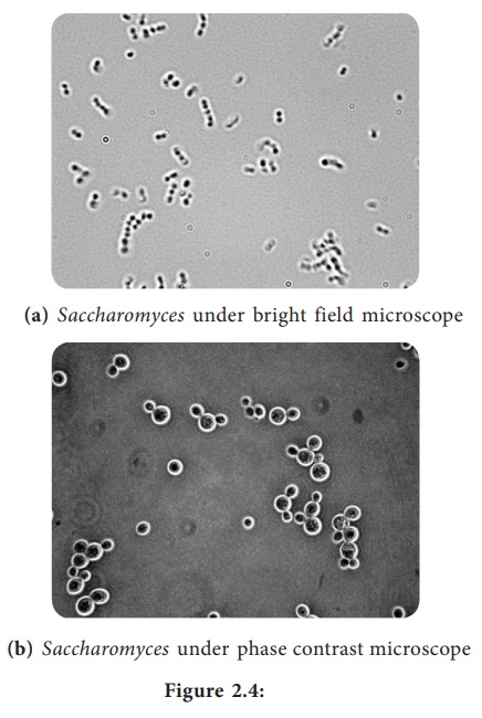
(Wavelength) gives the contrast for clear visibility of the object. Figure 2.4
Microscopic image comparing phase and bright field microscopy.
Infobits
Whenever light (or any wave in general) goes from one medium to
another, some of the energy of the wave is “reflected” back through the first
medium cut the same angle as the incident wave and some of the energy is
refracted (bent). Through the second medium when light goes from a low
refractive index medium to a high refractive index medium such as air to water
the reflection undergoes a 180° phase change. Light waves that are in phase
(that is, their peaks and valleys exactly coincide) reinforce one another and
their total intensity increases.
Light waves that are out of phase by exactly one-half wavelength
cancel each other and result in no intensity. That is darkness wavelengths that
are out of phase by any amount will produce some degree of cancellation and
result in brightness less than maximum, but more than darkness. Thus, contrast
is provided by differences is light intensity that result from differences in
refractive indices in parts of the specimen that put light waves indices in parts
of the specimen that put light waves more or less out of phase. As a result,
the specimen appears as various levels of darks against a bright background.
Applications
• Phase
contrast microscope enables the visualization of unstained living cells.
• It makes highly transparent objects more visible
• It is used to examine various intracellular
components of living cells at relatively high resolution.
• It helps in studying cellular events such as cell
division.
• It is
used to visualize all types of cellular movements such as chromosomal and
flagellar movements..
Related Topics