Chapter: 12th Microbiology : Chapter 2 : Microscopy
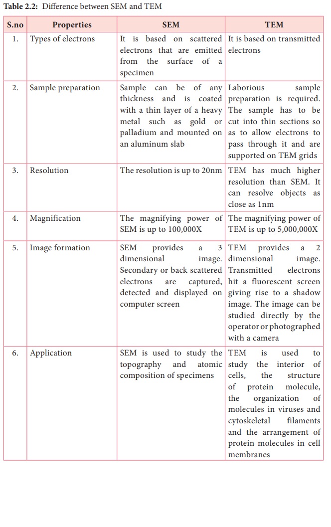
Difference between SEM and TEM
Difference
between SEM and TEM

SEM (Scanning electron microscopes)
1. Types of electrons : It is
based on scattered electrons that are emitted from the surface of a specimen
2. Sample preparation : Sample
can be of any thickness and is coated with a thin layer of a heavy metal such
as gold or palladium and mounted on an aluminum slab
3. Resolution : The resolution is up to 20nm
4. Magnification : The
magnifying power of SEM is up to 100,000X
5. Image formation : SEM
provides a 3 dimensional image. Secondary or back scattered electrons are
captured, detected and displayed on computer screen
6. Application : SEM is used to study the
topography and atomic composition of specimens
TEM (Transmission electron microscopes)
1. Types of electrons : It is
based on transmitted electrons
2. Sample preparation :
Laborious sample preparation is required. The sample has to be cut into thin
sections so as to allow electrons to pass through it and are supported on TEM
grids
3. Resolution : TEM has much higher resolution
than SEM. It can resolve objects as close as 1nm
4. Magnification : The
magnifying power of TEM is up to 5,000,000X
5. Image formation : TEM
provides a 2 dimensional image. Transmitted electrons hit a fluorescent screen
giving rise to a shadow image. The image can be studied directly by the
operator or photographed with a camera
6. Application : TEM is used to study the interior
of cells, the structure of protein molecule, the organization of molecules in
viruses and cytoskeletal filaments and the arrangement of protein molecules in
cell membranes
Related Topics