Principle, Working Mechanism, Applications, Optical Components - Fluorescence Microscope | 12th Microbiology : Chapter 2 : Microscopy
Chapter: 12th Microbiology : Chapter 2 : Microscopy
Fluorescence Microscope

Fluorescence Microscope
Fluorescence
microscope is a very powerful analytical tool that combines the magnifying
properties of light microscope with visualization of fluorescence.
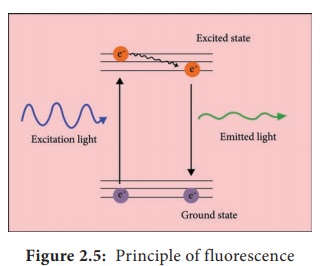
Fluorescence microscope is a type of light microscope which instead of utilizing visible light to illuminate specimens, uses a higher intensity (lower wavelength) light source that excites a fluorescent molecule called a fluorophore (also known as fluorochrome). Fluorescence is a phenomenon that takes place when the substances (fluorophore) absorbs light at a given wavelength and emits light at a higher wavelength. Thus, fluorescence microscopy combines the magnifyin properties of the light microscope with fluorescence technology.
Infobits
British scientist Sir George G. Stokes first described
fluorescence in 1852. He observed that the fluorophore emitted red light, when
it was illuminated by ultraviolet excitation. Stokes noted that fluorescence
emission always occurred at a longer wavelength than of the excitation light.
This shift towards longer wavelength is known as stokes shift.

The
fluorophore absorbs photons leading to electrons moving to a higher energy
state (excited state). When the electrons return to the ground state by losing
energy, the fluorophore emits light of a longer wavelength (Figure 2.5). Three
of the most common fluorophores used are Diamidino – phenylindole (DAPI) (emits
blue), Fluorescein isothiocyanate (FITC) (emits green), and Texas Red (emits
red).
Principle
Light
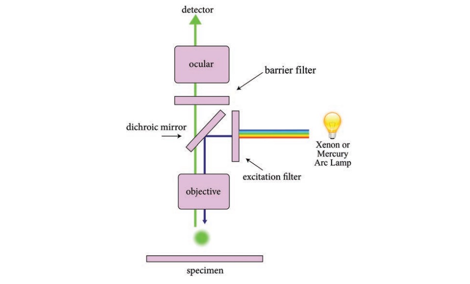
source such as Xenon or Mercury Arc Lamp which provides light in a wide range
of wavelength, from ultraviolet to the infrared is directed through an exciter
filter (selects the excitation wavelength). This light is reflected toward the
sample by a special mirror called a dichroic mirror, which is designed to
reflect light only at the excitation wavelength. The reflected light passes
through the objective where it is focused onto the fluorescent specimen. The
emissions from the specimen are in turn, passed back up through the objective
where magnification of the image occurs and through the dichroic mirror.
This
light is filtered by the barrier filter, which selects for the emission
wavelength and filters out contaminating light from the arc lamp or other
sources that are reflected off from the microscope components. Finally, the
filtered fluorescent emission is sent to a detector where the image can be
digitized.
Components of Fluorescence Microscope
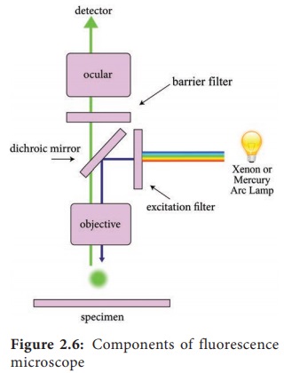
The main
components of the fluorescent microscope resemble the traditional light
microscope. However, the two main difference are the type of light source used
and the use of the specialized filter elements (Figure 2.6).

Light source
Fluorescence
microscopy requires a very powerful light source such as a Xenon or Mercury Arc
Lamp. The light emitted from the Mercury Arc Lamp 10–100 times brighter than
most incandescent lamps and provides light in a wide range of wavelengths from
ultra-violet to the infrared. Lasers or high-power LEDs were mostly used for
complex fluorescence microscopy techniques.
Filter elements
A typical
fluorescence microscope consists of three filters: excitation, emission and the
dichroic beam splitter.
Excitation filters: It is placed within the illumination path of a fluorescence microscope. Its purpose is to filter out all wavelength of the light source, except for the excitation range of the fluorophore in the sample or specimen of interest
Emission
filters: The emission filter is placed within the imaging path of a fluorescence
microscope. Its purpose is to filter out the entire excitation range and to
transmit the emission range of the fluorophore in the specimen.
Dichroic
filter or beam splitter: The dichroic filter or beam splitter is placed in
between the excitation filter and emission filter, at 45° angle. Its purpose is
to reflect the excitation wavelength towards the fluorophore in the specimen,
and to transmit the emission wavelength towards the detector.
Fluorescence is called “cold light” because it does not come
from a hot source like an incandescent light bulb.
Working Mechanism
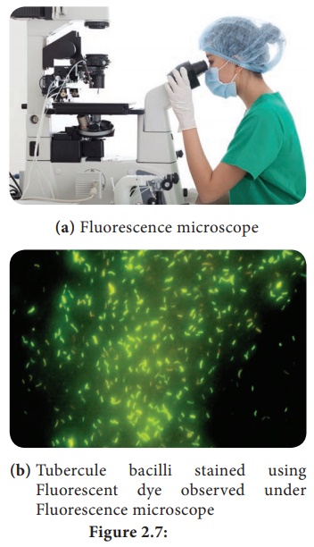
The specimen to be observed are stained or labeled with a fluorescent dye and then illuminated with high intensity ultra violet light from mercury arc lamp. The light passes through the exciter filter that allows only blue light to pass through. Then the blue light reaches dichroic mirror and reflected downward to the specimen. The specimen labeled with fluorescent dye absorbs blue light (shorter wavelength) and emits green light. The emitted green light goes upward and passes through dichroic mirror, reflects back blue light and allows only green light to pass the objective lens, then it reaches barrier filter which allows only green light. The filtered fluorescent emission is sent to a detector where the image can be digitized Figure 2.7.

Infobits
The Two Types of Fluorescence Microscopes includes diascopic
fluorescene and episcopic fluorescene.
Diascopic Fluorescence: K. Reichert and O.
Heimstadt demonstrated a fluorescence microscope using auto fluorescent
specimens in 1911.
This first type of fluorescence microscopy used transmitted
light. Light from the illumination source first passes through an excitor
filter and subsequently to the specimen through a dark field condenser. This
eliminates most of the excitation light from the imaging side of the system.
Episcopic Fluorescence: In episcopic fluorescence
microscopy, the excitation light comes from above the specimen through the
objective lens. This is the most common form of fluorescence microscopy today.
In this microscope, objective lens acts as both condenser and objective. Quartz
objective lenses are required for deep ultraviolet excitation.
Application
• Fluorescence microscope has become one of the most powerful techniques in biomedical research and clinical pathology.
• Fluorescence microscope allows the use of
multicolour staining, labeling of structures within cells, and the measurement
of the physiological state of a cell.
• Fluorescence microscope helps in observing
texture and structure of coal.
• To study porosity in ceramics, using a
fluorescent dye.
• To
identify the Mycobacterium tuberculosis.
Related Topics