Types, Principle, Working Mechanism, Applications, Instrumentation - Electron Microscope | 12th Microbiology : Chapter 2 : Microscopy
Chapter: 12th Microbiology : Chapter 2 : Microscopy
Electron Microscope
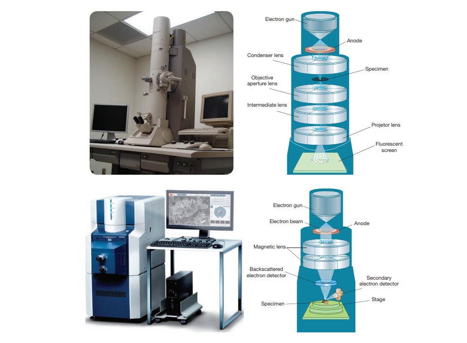
Electron Microscope
Examining
the ultra structure of cellular components such as nucleus, plasma membrane,
mitochondria and others requires 10,000X plus magnification which was just not
possible using Light Microscopes. This is achieved by Electron microscopes
which have greater resolving power than light microscopes and can obtain higher
magnifications.
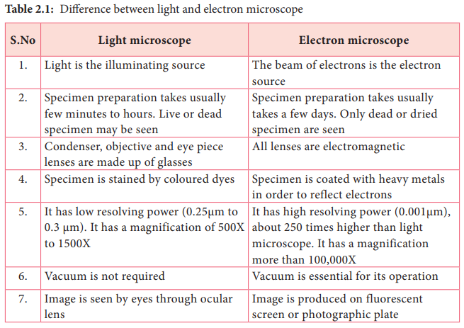
In an electron microscope, a focused electron beam is used instead of light to examine objects. Electrons are considered as radiation with wavelength in the range 0.001–0.01 nm compared to 400–700 nm wavelength of visible light used in an optical microscope. Optical microscopes have a maximum magnification power of 1000X, and resolution of 0.2 μm compared to resolving power of the electron microscope that can reach 1,000,000 times and resolution of 0.2 nm. Hence, electron microscopes deliver a more detailed and clear image compared to optical microscopes. Table 2.1 differentiate electron microscope from light microscope
(b) Tubercule bacilli stained using
Fluorescent dye observed under Fluorescence microscope
1924, a French sci-entist, Dr. De Brogli showed that an elec- tron
beam behaved like waves and had a wavelength much shorter than the sizes of
mole-cules and atom when accelerated.
Types of Electron Microscopes
• Transmission electron microscopes (TEM)
• Scanning
electron microscopes (SEM)
• Scanning transmission electron microscopes (STEM)
The
electron microscope was invented in 1931 by two German scientists, Ernst Ruska
and Max Knoll. Ernst Ruska later received Nobel Prize for his work in 1986. The
Transmission Electron Microscope (TEM) was the first type of Electron
Microscope to be developed
Principle
The
fundamental principle of electron microscope is similar to light microscope. In
electron microscope, a high velocity beam of electrons is used instead of
photons. In the electron gun, electrons are emitted from the surface of the
cathode and accelerated towards the anode by high voltage to form a high energy
electron beam. All lenses in the electron microscope are electromagnetic.
Charged electrons interact with the magnetic fields and magnetic force focuses
an electron beam. The condenser lens system controls the beam diameter and
convergence angles of the beam incident on a specimen. The image is formed
either by using the transmitted beam or by using the diffracted beam. The image
is magnified and focused onto an imaging device, such as a fluorescent screen,
on a layer of photographic film, or to be detected by a sensor.
Sample Preparation
Preparation
of specimens is the most complicated and skillful step in EM. The material to
be studied under electron microscopy must be well preserved, fixed, completely
dehydrated, ultrathin and impregnated with heavy metals that sharpen the
difference among various organelles.
The
material is preserved by fixation with glutaraldehyde and then with osmium
tetroxide. The fixed material is dehydrated and then embedded in plastic (epoxy
resin) and sectioned with the help of diamond or glass razor of
ultra-microtome.
In TEM,
sample sections are ultrathin about 50–100 nm thick. These sections are placed
on a copper grid and exposed to electron dense materials like lead acetate,
uranylacetate, phosphotungstate. In SEM, samples can be directly imaged by
mounting them on an aluminum stub.
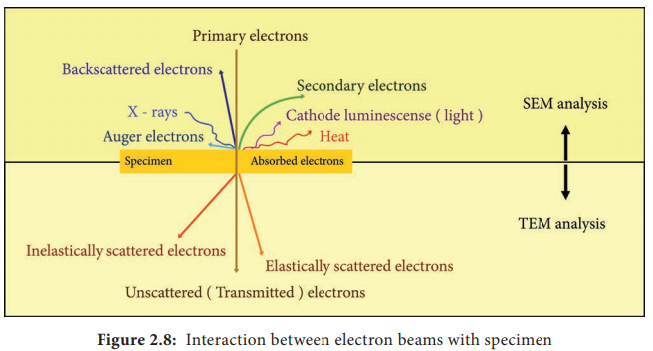
Electron–Sample Interactions
Interaction
of electron beam with the sample results in different types of electrons:
Elastic scattered electrons, Inelastic scattered electrons, secondary electrons
and backscattered electrons. Almost all types of electron interactions can be
used to retrieve information aboutthe specimen. Depending on the kind of
radiation or emitted electrons which are used for detection, different
properties of the specimen such as topography, elemental composition can be
concluded. Figure 2.8 shows the interaction of the electron beam with the
specimen
In
Transmission electron microscope (TEM), a beam of electrons is transmitted
through an ultrathin specimen, interacting with the specimen as it passes
through. An image is formed from the interaction of the transmitted unscattered
electrons through the specimen.
Secondary electrons are mainly used in scanning electron microscope (SEM) for imaging the surface topography of biological specimens. The interaction of electron beam with samples results in secondary electrons and backscattered electrons that are detected by standard SEM equipment
Working Principle and Instrumentation of TEM.
The
optics of the TEM is similar to conventional transmission light microscope.
A transmission
electron microscope has the following components,
1. Electron
gun
2. Condenser
lens
3. Specimen
stage
4. Objective
lens and projector lens
5. Screen/photographic
film/Charged Coupled Device (CCD) camera
Electron
Gun consists of a tungsten filament or cathode that emits electrons on
receiving high voltage electric current (50,000–100,000 volts). A high voltage
between the electron source (cathode) and an anode plate is applied leading to
an electrostatic field through which the electrons are accelerated
The emitted electrons travel through vacuum in the microscope column. Vacuum is essential to prevent strong scattering of electrons by gases. Electromagnetic condenser lenses focus the electrons into a very thin beam. Electron beam then travels through the specimen and then through the electromagnetic objective lenses. In a TEM microscope, the sample is located in the middle of the column. At the bottom of the microscope, unscattered electrons hit the fluorescent screen giving image of specimen with its different parts displayed in varied darkness, according to their density. The image can be studied directly, photographed or digitally recorded. Figure 2.9 show the arrangement of components for transmission electron microscope

Information
that can be obtained using TEM include,
• Topography: surface features, texture
• Morphology: shape and size of the particles
• Crystallographic arrangement of atoms
• Composition:
elements and the their relative amounts.
Working Principle and Instrumentation of SEM
It is
first built by Knoll in 1935. It is used to study the three dimensional images
of the surfaces of cells, tissues or particles. The SEM allows viewing the
surfaces of specimens without sectioning. The specimen is first fixed in liquid
propane at-180°C and then dehydrated in alcohol at-70°C. The dried specimen is
then coated with a thin film of heavy metal, such as platinum or gold, by
evaporation in a vacuum provides a reflecting surface of electrons. In SEMs,
samples are positioned at the bottom of the electron column and the scattered
electrons (back-scattered or secondary) are captured by electron detectors.
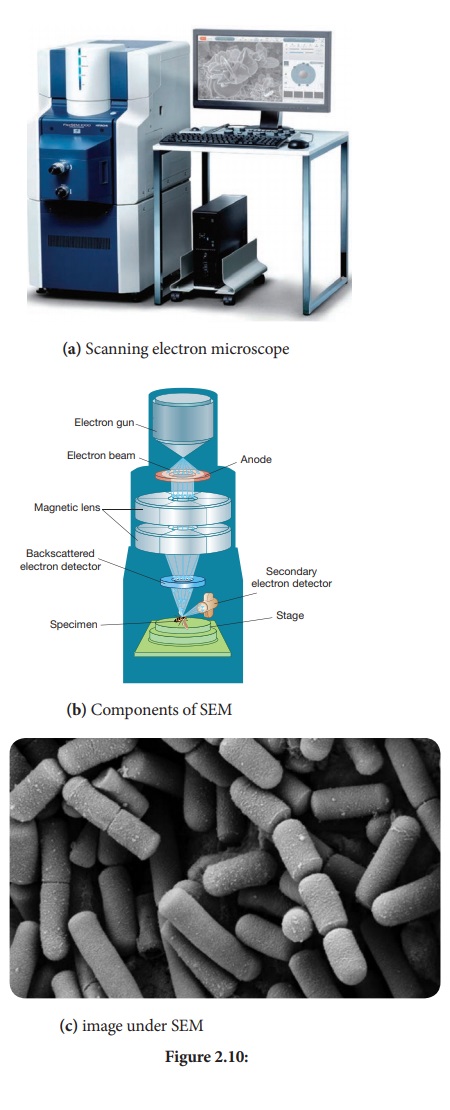
In SEM,
there are several electromagnetic lenses, including condenser lenses and one
objective lens. Electromagnetic lenses are for electron probe formation, not
for image formation directly, as in TEM. Two condenser lenses reduce the
crossover diameter of the electron beam. The objective lens further reduces the
cross-section of the electron beam and focuses the electron beam as probe on
the specimen surface (Figure 2.10). Objective lens thus functions like a
condenser. This is in contrast to TEM where objective lens does the
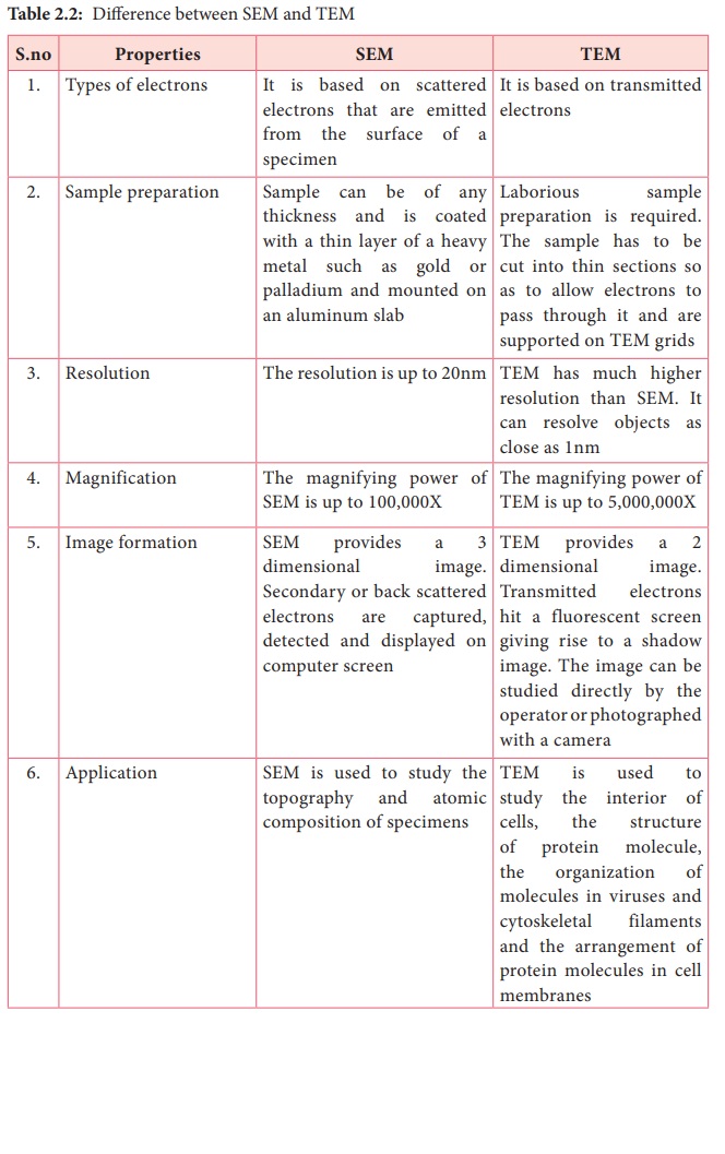
magnification. Major difference between SEM and TEM are given in Table 2.2.
SEMs are equipped with an energy dispersive spectrometer (EDS) detection system
which is able to detect and display most of the X-ray spectrum. The detector normally
consists of semiconducting silicon or germanium.
Scanning transmission electron microscopy (STEM) combines the principles of transmission electron microscopy and scanning electron microscopy and can be performed on either type of instrument. Like TEM, STEM requires very thin samples and the primary electron beam is transmitted by the sample. One of its principal advantages over TEM is in enabling the use of other of signals that cannot be spatially correlated in TEM, including secondary electrons, scattered beam electrons, characteristic X-rays, and electron energy loss.
Foldscope – origami based paper
microscope
A foldscope is an optica microscope that can be assembled from
simple components, including a sheet of paper and a lens. It was developed by
an Indian Manu Prakash. It consists of the following parts which are as
follows: Lens stage, sample stage, panning guide, ramp, lens and magnetic
cuppler. It has the magnification of 140X and maximum of 2400X
Related Topics