Chapter: Human Neuroanatomy(Fundamental and Clinical): Cranial Nerve Nuclei
Other Connections of Cranial Nerve Nuclei
Other Connections of Cranial Nerve Nuclei
1. The somatic efferent and special visceral efferent nuclei of cranial nerves supply striated (skeletal)muscle. Their connections are, therefore, similar to those of ventral horn cells of the spinal cord (Fig. 10.4).
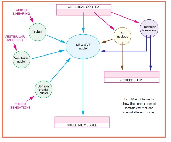
a. These nuclei are under cortical control through corticonuclear fibres. Cortical control of the hypoglossal nucleus is contralateral (i.e., from the opposite cerebral hemisphere). Corticonuclear fibres to the part of the facial nucleus innervating the lower part of the face is contralateral; but fibres for the rest of the nucleus are bilateral. The trochear nucleus receives ipsilateral fibres (i.e., from the same side). All other nuclei are influenced by both cerebral hemispheres, but the fibres to the abducent nerve are predominantly crossed. Corticonuclear fibres probably terminate by synapsing with interneurons present in, or near, the nuclei. Some corticonuclear fibres follow an aberrant path in the brainstem and travel through the medial lemniscus.
b. The nuclei are also influenced by other centres, namely the tectum, the red nucleus, and the reticular formation. The cerebellum, the diencephalon and the corpus striatum can influence the nuclei through these centres.
c. The motor nuclei also receive afferents from sensory nuclei of the brainstem including the nucleus solitarius and the spinal nucleus of the trigeminal nerve. The motor nucleus of the trigeminal nerve receives some fibres from the mesencephalic nucleus. These fibres form part of the pathway for the jaw jerk.
d. Motor nuclei may be connected to each other. The nuclei of the third, fourth and sixth cranial nerves are connected to each other, and to some other nuclei, through the medial longitudinal bundle. Fibres from the oculomotor, trochlear and abducent nuclei represent the final output of an elaborate system that controls eye movements.
2. The general visceral efferent nuclei innervatesmooth muscle and glands. Their central connections are less well known.
a. They receive afferents from sensory cranial nerve nuclei; in particular from the nucleus of the solitary tract.
b. Visceral sensations may also reach these nuclei through collaterals from ascending tracts. The exact pathways are not known.
c. The nuclei are connected with the reticular formation. Higher control is probably exercised by the hypothalamus. The cerebral cortex (specially the limbic lobe) and the thalamus may influence the nuclei through the hypothalamus.
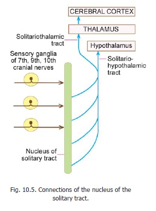
3. The nucleus of the solitary tract (GVA, SVA)receives afferents from the viscera supplied by nerves connected to it. The nucleus also receives fibres from the cerebral cortex and from the cerebellum. These afferents may modify the transmission of sensory impulses.Fibres arising in this nucleus carry visceral impulses to the hypothalamus and the thalamus through the solitario-hypothalamic and solitario-thalamic tracts respectively. Thenucleus of the solitary tract also sends fibres to the reticular formation, the general visceral efferent nuclei and to the spinal cord.
4. The main afferents of the nuclei of the generalsomatic afferent column (made up of the main and spinal nuclei of the trigeminal nerve) are central processes of neurons in the trigeminal ganglion. After entering the pons, many of these processes divide into ascending and descending branches; while others ascend or descend. The ascending fibres end in the main sensory nucleus. Those that descend form a large bundle of fibres that constitutes the spinal tract of the trigeminal nerve (Fig. 10.6). The tract is closely related to the dorsolateralaspect of the spinal nucleus of the trigeminal nerve. The tract descends from the pons into the medulla, and then into the upper part of the spinal cord. (Apart from trigeminal fibres the tract also carries general somatic afferent fibres from the facial, glossopharyngeal and vagus nerves).
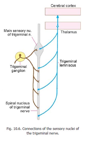
The fibres ending in the main sensory nucleus are concerned, predominantly, with touch; and those ending in the spinal nucleus are concerned, predominantly, with sensations of pain and temperature. However, the functional distinction between the two nuclei is by no means clear cut. These nuclei also receive descending fibres from the cerebral cortex: these fibres may influence the conduction of sensory impulses.
The lowest part of the spinal nucleus of the trigeminal nerve (nucleus caudalis) is of particular importance in pain reception. It receives nerve fibres from the nucleus raphe magnus that modulate pain transmission. (These nerve fibres contain enkephalin, noradrenaline and serotonin).
The neurons in the main sensory and spinal nuclei of the trigeminal nerve are second order neurons. Their axons are comparable to the fibres of the spinothalamic tracts. The axons cross to the opposite side and form a bundle called the trigeminal lemniscus (Fig. 10.6). This lemniscus ascends to the thalamus (ventral posteromedial nucleus). Some fibres in the lemniscus may be uncrossed. Third order neurons located in the thalamus carry the sensations to the sensory areas of the cerebral cortex. In the pons and midbrain the trigeminal lemniscus lies immediately lateral to the medial lemniscus (Figs. 11.5, 11.7 to 11.9). Its fibres are closely related to those of the spinal lemniscus (lateral spinothalamic tract).
A separate bundle of more dorsally situated trigeminothalamic fibres is also described.
In addition to the efferents to the thalamus, the trigeminal nuclei probably send fibres to other cranial nerve nuclei, reticular formation, cerebellum, and hypothalamus.
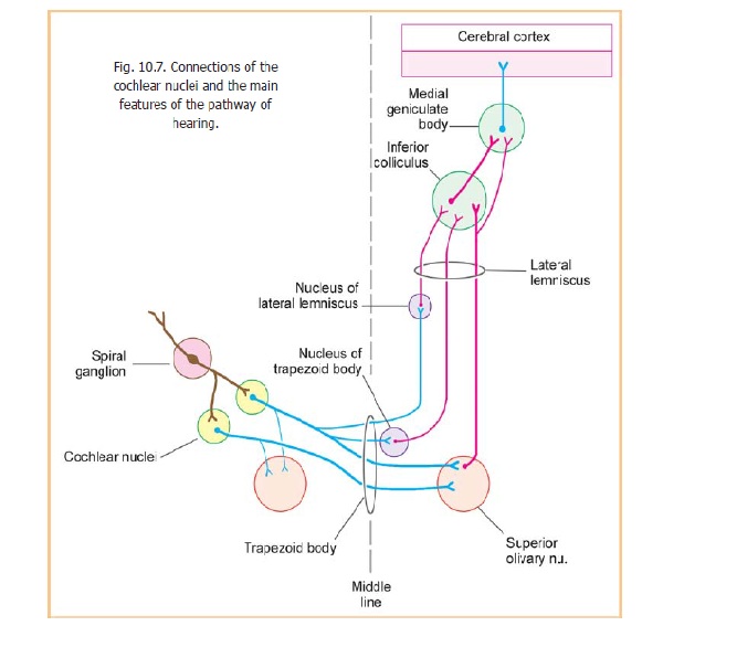
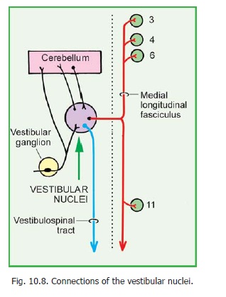
5. The special somatic afferent nuclei (cochlear and vestibular) have important connections (Figs.10.7, 10.8).
Related Topics